The auricular branch of the vagus or Arnold’s nerve is also called the Alderman’s nerve. The term arose because it was alleged that voracious aldermen and their guests, during massive banquets, would scratch their outer ears to evoke vomiting [1] and thereby relieve their over-distended stomachs to permit yet further gluttony. This perhaps is the modern equivalent of the Romans’ mythical visit to the vomitorium between courses.
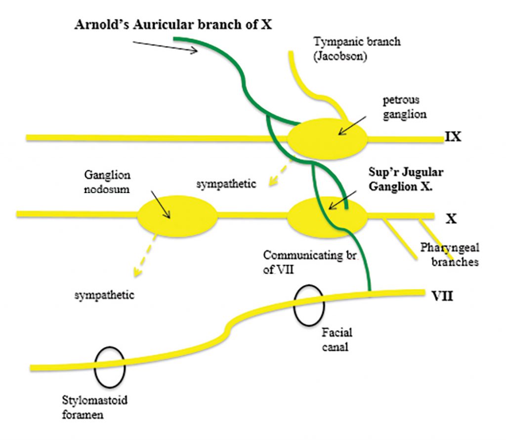
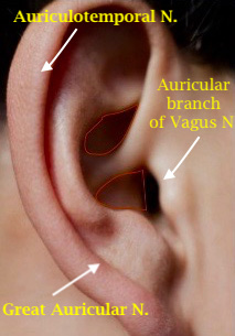
Arnold’s nerve conveys sensation from the tragus and external acoustic meatus (Figures 1 & 2). It is a mixed general somatic afferent nerve composed of vagal, glossopharyngeal, and facial nerve fibres. Confusingly, the term is also applied to the greater occipital nerve of Arnold [2]. The eponym has been associated with the related Arnold’s canal— the passage in the petrous temporal bone for Arnold’s nerve, and Arnold’s otic ganglion.


Arnold’s nerve is the remnant of the embryonic nerve that supplies the first branchial arch, which includes the external acoustic meatus, middle ear and auditory tube. The auricular nerve arises from the superior jugular ganglion of the vagus [3] and is joined by a filament from the inferior (petrous) ganglion of the glossopharyngeal (Figure 1). Crossing the temporal bone about 4mm above the stylomastoid foramen, it gives off an ascending branch that joins the facial nerve. Fibres of the nervus intermedius of Wrisberg (VII) may join the auricular nerve explaining the vesicles that sometimes accompany geniculate zoster.
In original studies of comparative anatomy in 1913-22 [4,5], the eminent, Glasgow Otologist Albert A Gray (1869-1936) elucidated the minutiae of anatomical detail and its variations in both man and animals, and described the ‘bullar plexus’. He noted that descriptions of the course and relations of Arnold’s nerve ‘in standard anatomical textbooks were quite inadequate and probably incorrect.’ He demonstrated:
…. This plexus was clearly composed of branches from at least two nerves, the facial and the vagus; and probably branches from the glossopharyngeal also took part in its formation. The existence of this structure in several mammals of different orders appeared to me to indicate that it was probably represented in man, but had hitherto escaped discovery on account of the difficulties of making satisfactory dissection of that region in the human subject…
Arnold’s nerve enters the temporal bone on the external surface of the jugular fossa, and runs horizontally where a small twig unites it with the facial nerve. Turning round the posterior aspect of the facial nerve it comes to be immediately behind the chorda tympani with which it connects, then turns more abruptly downwards and leaves the bone through a small foramen close to the style-mastoid foramen…The bullar plexus indeed in the human subject consists of the communications of Arnold’s nerve (the auricular branch of the vagus) with the facial nerve, and in some cases at least with the chorda tympani [5].
Gray stressed the differences between mammals and man in whom ‘the development of the mastoid antrum and the mastoid cells compels the facial nerve to seek a more devious course [4].
Clinical
Because Arnold’s nerve innervates the external auditory canal, when the canal is stimulated by scratching, the reflex vagal response is vomiting, or a cough – that is Arnold’s ear-cough reflex [6]. The afferent projection from the auricular branch to the nucleus tractus solitarius connects it to the efferents in the dorsal nucleus of the vagus nerve and the nucleus ambiguus which supply inter alia the heart and lungs. Arnold’s nerve has also been claimed as the basis for so-called auriculo-palatal, auriculo-lacrimal, auriculo-cardiac reflexes, which are of minor clinical significance [6].
Gupta et al. reported a survey of 500 patients that revealed a 4.2% incidence of Arnold’s ear-cough reflex [6]. In another clinical survey of 688 patients an incidence of 1.74% was found, usually unilaterally, and elicited from each canal quadrant [7]. A positive reflex did not indicate any particular ear disease. Arnold’s reflex has been claimed as an occasional cause of chronic cough in children and adults [8] with earwax and in those wearing hearing aids. There is however a danger of over-diagnosis of this syndrome in practice, as too easy an explanation for chronic cough which should necessitate careful investigation.

“The National Library of Medicine believes this item to be in the public domain.”
The nerve may be involved by glomus jugulare and other tumours, and by trauma involving the lateral posterior fossa and jugular canal. Autosomal dominant hereditary sensory neuropathy HSN1 can rarely present with adult onset paroxysmal cough triggered by noxious odours or by pressure in the external auditory canal (Arnold’s ear-cough reflex), gastro-oesophageal reflux, a hoarse voice, cough syncope and sensorineural hearing loss [9].
Friedrich Arnold (1803 – 1890) (Figure 3) studied anatomy at the University of Heidelberg under Friedrich Tiedemann (1781-1861). He graduated in Medicine in September 1825. In 1834 he was appointed Extraordinary Professor, Faculty of Medicine, at Heidelberg, and in 1835 became Professor of Anatomy at the University of Zurich.
Arnold described the external arcuate fibres (Arnold’s bundle), the arcuate nuclei and the otic ganglion (Arnold’s ganglion). In his Tabulae Anatomicae quas ad Naturam Accurate Descriptas he first depicted the frontopontine tract from the frontal cortex through the anterior limb of the internal capsule via the cerebral peduncle (Arnold’s tract). Arnold himself noted the reflex cough when the external auditory meatus was stimulated.
Of many publications on microanatomy and physiology of the nervous system, his textbook, published with his brother Johann Wilhelm Arnold (1801–1873) [10], was the most respected. His Icones nervorum capitis (1834) [11], provided lithographic engravings of the cranial nerves showing their topography, ‘with a distinctness and beauty never been seen before [12]’. He was regarded as one of the greatest dissectors of all time.
His pupils included Ludwig Edinger (1855-1918) and Hubert von Luschka (1820–1875). He moved again in 1845 to the Chair at Tübingen, and finally returned to his original Chair at Heidelberg, where he died aged 87.
References
- Treves F. Surgical Applied Anatomy. London: Cassell & Co. 1883.
- Vital JM, Grenier F, Dautheribes M, Baspeyre H, Lavignolle B, Sénégas J. An anatomic and dynamic study of the greater occipital nerve (N. of Arnold). Surg Radiol Anat. 1989;11(3):205-10. https://doi.org/10.1007/BF02337823
- Tekdemir I, Aslan A, Elhan AA. clinico-anatomic study of the auricular branch of the vagus nerve and Arnold’s ear-cough reflex Surg Radiol Anat. 1998;20:253-7. https://doi.org/10.1007/s00276-998-0253-5
- Gray AA. Comparative Anatomy of the Middle Ear. Journal of Anatomy and Physiology 1913;47:391-413.
- Gray AA. The Course and Relations of Arnold’s Nerve (Auricular Branch of the Vagus) in the Temporal Bone. Proc R Soc Med. 1922; 15(Otol Sect):15-18. https://doi.org/10.1177/003591572201501115
- Gupta D, Verma S, Vishwakarma SK. Anatomic basis of Arnold’s ear-cough reflex. Surg Radiol Anat. 1986;8:217-20. https://doi.org/10.1007/BF02425070
- Bloustine S, Langston L, Miller T. Ear-cough (Arnold’s) reflex. Ann Otol Rhinol Laryngol. 1976;85:406-7. https://doi.org/10.1177/000348947608500315
- Jegoux F, Legent F, de Montreuil C. Chronic cough and ear wax . Lancet. 2002;360:618. https://doi.org/10.1016/S0140-6736(02)09786-6
- Spring, PJ, Kok, C, Nicholson, GA, Ing, AJ. et al. Autosomal dominant hereditary sensory neuropathy with chronic cough and gastro-oesophageal reflux: clinical features in two families linked to chromosome 3p22-p24. D. Brain 2005;128 :2797-2810 https://doi.org/10.1093/brain/awh653
- Arnold F, Arnold JW. Die Erscheinungen und Gesetze des lebenden menschlichen Körpers im gesunden und kranken Zustande. [on the phenomena and laws of the living human body in the healthy and diseased state] 2 volumes. Zürich. Orell Füssli, Verlag. , 1836-1837.
- Arnold F. Icones cerebri et medullae spinalis. Orell Füssli, Verlag. 1838.
- Hildebrand R. The cranial nerves as living morphotic entities- Friedrich Arnold’s Icones Nervorum Capitis, Heidelberg 1834. Acta Neurochir (Wien) 1988;92:5-14.
https://doi.org/10.1007/BF01401966