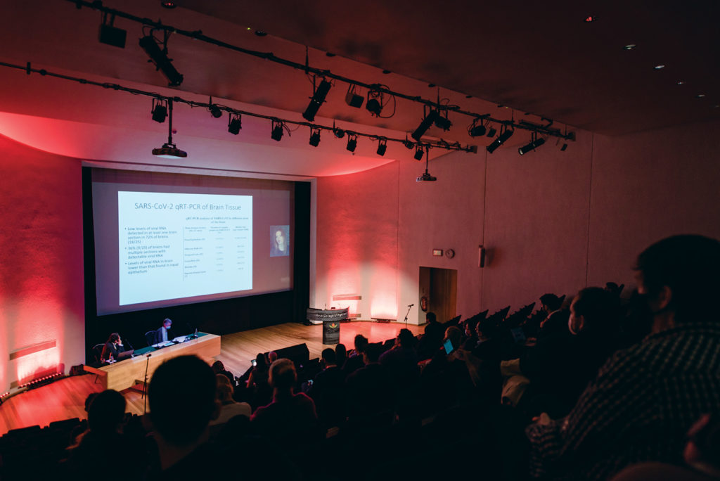
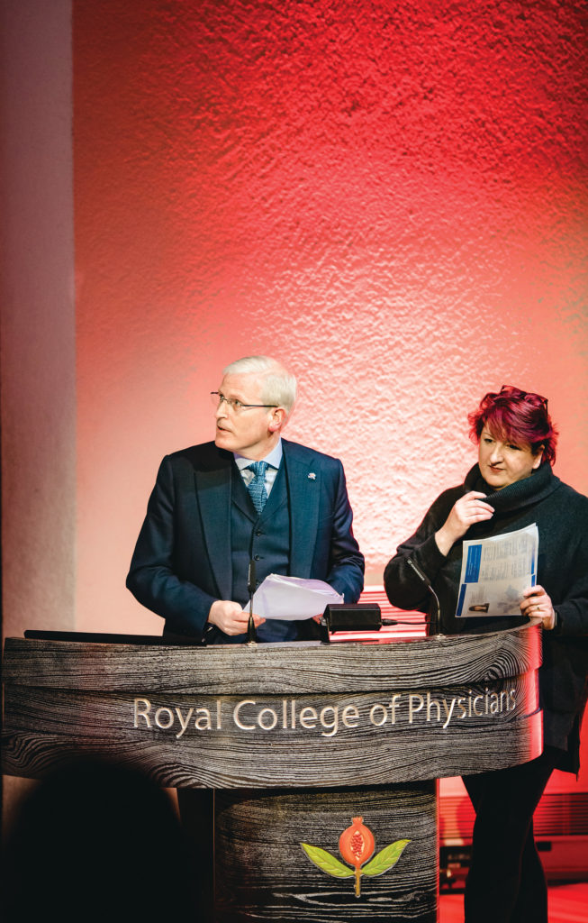
On December 7th, 2021, the Encephalitis Conference successfully took place in a hybrid format despite the COVID-19 pandemic. The conference was delivered to 314 delegates in-person and digitally from 50 countries, welcoming both researchers and healthcare professionals worldwide with an interest in a broad range of subjects related to encephalitis.

The conference began with a session chaired by Professor Angela Vincent, Emeritus Professor of Neuroimmunology at the University of Oxford, UK and moderated by Dr Benedict Michael, Vice Chair of the Society’s Scientific Panel and Senior Clinician Scientist Fellow at the University of Liverpool, UK.
Dr Helena Ariño, Institute of Psychiatry, Psychology and Neuroscience, King’s College, London, UK, presented on how clinical texts from electronic health records (EHRs) could be used to diagnose anti-NMDAR encephalitis. She discussed how anti-NMDAR encephalitis, which typically presents with prominent psychiatric symptoms, could mimic a primary psychiatric disorder leading to delays in diagnosis and treatment. Her results suggest that clinical text contained within EHRs can contain information which can aid in the diagnosis of anti-NMDAR encephalitis and its differentiation from other phenotypically similar conditions. These findings have important clinical relevance.
Dr Emily Happy Miller, Columbia University Irving Medical Centre and the New York Presbyterian Hospital, USA, presented her work exploring the neuroinvasive potential of SARS-CoV-2 in patients previously infected with SARS-CoV-2 who had died and undergone autopsy. Dr Miller reported that the abnormal pathological findings found in the autopsied brains were likely due to systemic inflammation rather than direct viral invasion of the brain parenchyma tissue by SARS-CoV-2. This demonstrates an important step in understanding what may be occurring in the brains in those representing a more severe phenotype of SARS-CoV-2.
Professor Virginie Desestret, Hospices Civils de Lyon and Université Claude Bernard in Lyon, France explored the mechanisms in which breast carcinomas can trigger Yo paraneoplastic cerebellar degeneration (PCD). Yo-PCD is caused by autoimmunity against neural antigens expressed by tumour cells. They found that Her2 – hormone receptor and CDR2L, the main Yo antigen, likely had a pivotal role in breakdown of immune tolerance alongside several other biomolecular hallmarks. An understanding of these pathological mechanisms informs potential therapeutic targets.
The first keynote lecture was given by Professor Jerome Honnorat, Chair of Department of Neuro-Oncology at Hospices Civils de Lyon, France who presented on paraneoplastic encephalitis. He started by describing the association between autoantibodies and cancer and the classical paraneoplastic neurological syndromes (PNS). Subsequently, the associations between the closely related conditions of autoimmune encephalitis and PNS encephalitis were discussed, and their different pathophysiologies described. Professor Honnorat highlighted the need for the description of new syndromes and a better understanding of the incidence of paraneoplastic encephalitis. He also highlighted the importance of classifying the disease aetiology through antibodies, tumour markers and presence of cancer alongside the clinical symptoms and the need for new and better codified treatments.

After a short break the second morning session was chaired by Professor Sarosh Irani, Clinician Scientist and Consultant Neurologist at the John Radcliffe Hospital, Oxford, UK and moderated by Dr Thomas Pollak, NIHR Clinical Lecturer at the Institute of Psychiatry, Psychology and Neuroscience at King’s College London, UK.
Dr Bo Sun, Autoimmune Neurology Group, University of Oxford, UK described how an in vitro assay was used to examine the autoreactivity of CASPR2-autoantibodies across B-cell developmental stages. This work elucidates the role that late B-cell tolerance dysfunction may have in disease pathogenesis. This could aid in improving and developing novel therapeutic strategies for the long-term prevention of autoantibody production.
Dr Tina Damodar, Department of Neurovirology at NIMHANS, Bangalore, India focused on the identification of clinical, haematological and biochemical markers associated with acute encephalitis syndrome (AES) following rickettsial infection in children. An aetiology for AES was identified in 119 (55%) of the 216 children included in the study with 60 cases diagnosed as rickettsial encephalitis (scrub typhus and other rickettsial pathogens). Preliminary findings revealed that patients with rickettsial infection were characterised by specific biomarkers. This could lead to a potential diagnostic tool for rickettsial and other treatable causes of AES. Further studies involving larger sample sizes will inform on the generalisability of these findings.
Dr Jamil Kahwagi, Centre Hospitalier National Universitaire de Fann, Dakar, Senegal, presented an observational study exploring whether SARS-CoV-2 contributed to encephalitis and related illnesses in Dakar, Senegal. Within this observational study, 59 patients were enrolled. A viral aetiological agent was confirmed in 12 patients and a probable cause was found in 8 patients with SARS-CoV-2 being associated as the cause in six of these patients. These results show that SARS-CoV-2 has been detected in patients with encephalitis and related illnesses where no other aetiological agent is found. However, with only 34% of patients having an aetiological agent detected, this highlights the need for better diagnostic tools and greater surveillance of encephalitis in Senegal.
Dr Adam Al-Diwani and colleagues, University of Oxford, UK explored the role of germinal centre reactions in NMDA receptor-antibody encephalitis. In patients with an ovarian teratoma, they found evidence of these reactions in both blood and teratoma material itself (cultures, histology, and single cell sequencing). However, germinal centre reactions usually occur in a specialised system called lymphoid tissue. Working with expert radiologists, they used ultrasound to guide sampling of neck lymph nodes in patient volunteers. For the first time, they showed that some patients have NMDAR-producing cells in this region; but not in patients who had had a completely excised ovarian teratoma. The team conclude that germinal centre reactions are relevant to this illness – providing a basis for a thorough teratoma search and prompt removal if found, and beyond this, consideration of earlier B cell depleting treatments in patients without a teratoma.
Invited guest speaker, Assistant Professor Deanna Saylor from The Johns Hopkins University School of Medicine, USA described her experience in developing neurological care and training in resource-limited settings. Professor Saylor discussed the high burden of neurological disorders globally and in lower middle income countries and highlighted that with an ageing and growing population these problems are only set to increase. Moreover, the disparities in postgraduate neurology training globally were stark, with opportunities being absent from many lower middle income countries. Through her experiences in creating a neurology training programme in Zambia, a lower middle income country, she demonstrated how the lack of human and physical resources can make developing a system of neurological care challenging. However, through her work she has shown, despite these challenges, that with an on-the-ground leader and in-person clinical training it is possible to create an effective neurological programme and improve care.
After lunch, the first afternoon session was also hosted by Professor Sarosh Irani and moderated by Dr Jess Fish, Clinical Psychologist and Lecturer in Neuropsychological Rehabilitation at the Institute of Health and Wellbeing, University of Glasgow, UK.
The first speaker of the afternoon was Dr Jie Chen, Rockefeller University, New York, USA. Dr Chen’s presentation explored the pathogenesis of enterovirus rhombencephalitis in children. Whole exome sequencing was carried out on 15 unrelated patients with enterovirus rhombencephalitis and their parents. This elucidated the role that both human toll-like receptor 3 and melanoma differentiation-associated protein 5 have in intrinsic immunity to enterovirus rhombencephalitis through the control of baseline or virus-induced type 1 interferon (INF) production. The discovery of the role these receptors play could lead to new therapeutics therapies to treat patients with enterovirus rhombencephalitis such as IFN-α2b in early stage.
Ms Selina Yogeshwar, Charité–Universitätsmedizin, Berlin, Germany described how the research group conducted the first longitudinal study of brain atrophy patterns in anti-IgLON5 disease, using advanced magnetic resonance imaging, to better understand the underlying pathophysiological mechanisms of anti-IgLON5. They found that several patients had brainstem atrophy with different levels of progression. These results contribute to a better understanding of IgLON5 disease progression and differential longitudinal patterns. In the future, they seek to further include key co-variates such as age, sex, and disease stage to attain a more holistic and thorough picture of the neurodegeneration that accompanies anti-IgLON5 disease, while adjusting for important confounding variables.
The work presented by Dr Renata Barbosa Paolilo, HCFMUSP, São Paulo, Brazil, focused on relevant characteristics of encephalitis in children in a Brazilian tertiary centre and how these characteristics can change in the presence/absence of anti-NMDAR-antibodies. Through a retrospective analysis of these characteristics, they found that autoimmune encephalitis was the most common diagnosis of encephalitis, with most having the anti-NMDAR-antibody phenotype. Moreover, those with the anti-NMDAR-antibody phenotype had a more severe and polysymptomatic presentation and a more aggressive treatment approach was needed. The work demonstrates clinical differences in treating different types of encephalitis and highlights that more research is needed to evaluate anti-NMDAR-antibodies.
Dr Sukhvir Wright, Institute of Health and Neurodevelopment at Aston University, Birmingham, presented on a novel method of treating anti-NMDAR-Ab encephalitis through using the neuronal steroid pregnenolone sulphate (PregS) to upregulate anti-NMDAR function. They used an in vivo and vitro animal model involving rats and injected them with either control antibodies or human-derived anti-NMDAR-Ab. In vitro, they found that PregS resulted in an increase in anti-NMDAR function likely due to enhanced NMDAR trafficking at the surface of neuronal membranes. These results were also mimicked when PregS was applied in vitro to human brain tissue, supporting its potential efficacy as a future treatment of anti-NMDAR-Ab encephalitis.
The final session was chaired by Professor Tom Solomon, President of the Encephalitis Society and head of the Brain Infection Group, Liverpool, UK and moderated by Dr Jess Fish.
The final session of the day was Dr Álvaro Bonelli, Rey Juan Carlos University Hospital, Madrid, Spain. Dr Bonelli presented on how multiplex-PCR could be used to diagnose central nervous system infections (CNSIs). Dr Bonelli discussed how the delays in the diagnosis and treatment of CNSIs can lead to a high mortality and morbidity with traditional diagnostic methods identifying an aetiological agent in less than 40% of cases. When replacing traditional methods with multiplex-PCR, they found a significant increase in the proportion of patients where a causative agent was found. As such, by using multiplex-PCR both the diagnostic rate and treatment could be improved.
Dr Harschnitz, Sloan-Kettering Institute for Cancer Research, New York, USA, and Human Technopole, Milan, Italy, presented on how their group used human pluripotent stem cells (hPSCs) to investigate which neural cells are permissive to infection by SARS-CoV-2. Using hPSCs in both in vitro and in vivo models, they discovered that dopaminergic neurons are permissive to SARS-CoV-2 infection. This finding was mirrored in human autopsy samples which showed viral reads of SARS-CoV-2 in the midbrain and a loss of dopamine neurons. This work highlights dopaminergic neurones as a possible target for SARS-CoV-2 infection which triggers inflammation and cellular senescence.
The work presented by Adawa Manuela, COVID 19 ORCA Patient Management Center, Yaoundé, Cameroon presented on the aetiologies of encephalitis in non-HIV infected patients in Cameroon. After a screening process, 18 non-HIV infected patients, mostly men from rural areas, were included in this study. They found that the most frequent clinical signs were fever and disturbance of consciousness with the most common aetiological agent being Streptococcus Pneumoniae. This implies that Streptococcus Pneumoniae could be a common cause of encephalitis in non-HIV infected patients in Cameroon.
Charly Billaud, School of Health and Life Sciences at Aston University, Birmingham, UK. presented on the long-term neurophysiological outcomes in paediatric autoimmune encephalitis using magnetoencephalography (MEG). Whilst still an ongoing study, primary results in five children with autoimmune encephalitis suggest that the latency of recorded brain auditory responses is related to performance in working memory and processing speed. There are plans to expand this research to a larger cohort and dissect potential indicators of thinking skills.
The second keynote lecture was given by Winifred Mercer Pitkin and Assistant Professor Kiran Thakur from Columbia University Irving Medical Center, USA. Professor Thakur presented how arthropod-borne encephalitides are becoming an increasing problem globally through anthropomorphic factors such as encroachment onto vector habitats and global warming. This has significantly increased the area in which vectors associated with arthropod encephalitides can thrive. Furthermore, she described how many pathogens transmitted by ticks and mosquitos have common mechanistic pathways. Moreover, common host and pathogen immunogenetic factors have been associated with neurovirulence caused by these pathogens. This could pave the way for new potential therapeutic targets; however, more studies are needed to understand these immune pathways responsible for neuropathology.
Dr Ava Easton, CEO of the Encephalitis Society and Honorary Fellow, Dept. Clinical Infection, Microbiology and Immunology at the University of Liverpool began to close the conference with a video summarising how the Encephalitis Society has adapted to the COVID-19 pandemic and showcased events that took place in the past year despite this.

Dr Nicholas Davies and Dr Ava Easton presented the awards and prizes for best poster and best oral presentations:
Best poster for “The clinical diversity of anti-IgLON5 disease in the Dutch population” to Ms Yvette S Crijnen, from the Department of Neurology, Erasmus University Medical Center, Rotterdam, The Netherlands, (with colleagues Juliette Brenner, Inga Koneczny, Verena Endmayr, Catarina Alcarva, Nadine van der Beek, Chiara Glen, Ece Erdag, Daniëlle Bastiaansen, Agnita Boon).
Best oral presentation for “Dissecting CASPR2-antibody encephalitis with patient derived CASPR2-specific monoclonal antibodies” to Dr Bo Sun, from the University of Oxford/John Radcliffe Hospital, Oxford, UK and for “Encephalitis and autoimmune encephalitis in paediatric patients from Brazil” to Dr Renata Barbosa Paolilo from the Hospital das Clínicas da Faculdade de Medicina da Universidade de São Paulo (HCFMUSP), São Paulo, Brazil.
The conference concluded with a thanks to the conference sponsors and a closing call to action from Dr Ava Easton and Dr Nicholas Davies to get involved with World Encephalitis Day which was on 22nd February 2022 (www.worldencephalitisday.org).
The next Encephalitis Conference will be 30th November and 1st December 2022 at the Royal College Physicians. Sign up for free professional membership to be alerted to the program, and other opportunities such as seedfunding, grants, and bursaries: https://www.encephalitis.info/professional-membership