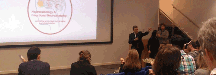
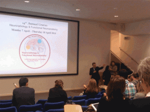
Organisers: Professor Thomas Naidich (Neuroradiology Mount Sinai, New York), Professor Christopher Yeo (Behavioural Neuroscience, UCL) and Professor Tarek Yousry (Lysholm Department of Neuroradiology, Queen Square).
Introduction
This four-day course required the attendees to familiarize themselves with functional anatomy, cytoarchitectonic organisation of the brain and its clinical relevance. Participants were able to apply the theoretical knowledge acquired and apply it to clinical practice through using interactive medical imaging and hands-on dissection workshops. The attendees were of varying expertise, with levels ranging from neuroscientists, neurosurgeons and neuroradiologists to neurology trainees like myself. Therefore, the course design accommodated non-clinicians and clinicians alike and coupled neuroscience with clinical neurology.
I chose to attend this course of interactive lectures delivered by world-class experts at the start of my specialist training to enhance my understanding of functional neuroanatomy.
Course contents
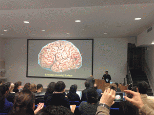
The course opened with an interesting insight into the phylogenetic evolution of the brain in humanoids, architectonic organisation of the telencephalon, neuroimaging with pre- and postnatal development of the white matter. It explored the fundamental functions of the brain in more depth such as the neurosignalling association pathways using functional imaging and tractography. Key regions covered were the cerebellum, corpus callosum, basal ganglia, hippocampus, amygdala, limbic system, vestibular system, insular and visual cortex.
Interesting learning points included
- Approximately 20% of population have bilateral symmetrical connections responsible for language (with women more likely to be in this group)
- The left arcuate fasiculus is larger than the right and therefore has a better chance of recovery following an insult.
- Being able to locate areas according to gyri for example Heschels Gyrus (Primary Auditory Cortex) located on the superior temporal gyrus.
Interactive components
The famous neuroanatomist Professor Naidich delivered interactive sessions with participants shouting key landmarks of the gyri and sulci and demonstrated how we could use our hands to make signs which guided us in recognition of key landmarks. His key landmark was the sylvian fissure and from there we could locate neighbouring structures. This required complete participation and was the most I had ever engaged in a course as it used different aids to help us recognize various structures. For instance demonstrating to us where the central sulcus is located and how it divides the frontal lobe from the parietal lobe on different planes on MRI.
We had anatomy workshop demonstrations, located at a nearby university, with specimen reviews and dissections in small groups. It was led by Professors Naidich and Yeo. This complemented the previous lectures and started with surface anatomy, identification of important landmarks i.e. sylvian fissure and proceeded on to discovering and locating deep brain structures in various different planes. The workshops were a fantastic opportunity to gain practical experience with dissected human brain sections and we had to identify key areas using information given in the earlier lectures.
The sessions that I had found most clinically relevant were the small group imaging sessions where we had to locate different brain regions systematically and identify the abnormality and its clinical relevance under the supervision of neuroradiologists. A mixture of pathologies were presented, ranging from tumours and stroke to neuromigrational abnormalities.
Summary
In summary this course is highly recommended for the neuroanatomy naïve professionals who require a working knowledge of neuroanatomy and an understanding of the relative functional and radiological significance of key brain regions.
The course was well organised, interactive and practical. It highlighted the research advances involved in improving our understanding of function, associating pathways and the clinical significance of key brain regions.
The 15th Annual Neuroradiology and Functional Neuroanatomy course takes place from the 23rd – 26th March 2015.
ACNR 2015;14(6);27. Published online 15/12/14