Abstract
The initial diagnosis as well as the long term management of occult spinal dysraphism and tethered spinal cord is often managed by a large number of healthcare professionals including Paediatricians, GPs, Neurologists, Neurosurgeons, Rehabilitation Physicians and Therapists. We review the entity of spinal dysraphism. An approach to the evaluation and diagnosis of these entities is subsequently discussed. In addition, concepts involved in the pathophysiology, neurosurgical repair, and outcome are presented in the context of postoperative management issues that rely upon the knowledge of all professionals who may encounter these patients.
Summary
- Embryology of spinal dysraphism
- Clinical features of tethered cord syndrome
- Multidisciplinary management of closed spinal dysraphism
Introduction
The finding of a midline spinal anomaly in a child typically prompts referral to the Paediatric Neurosurgeon for the evaluation of an occult spinal dysraphic state. Unfortunately, occult spinal dysraphism is not always readily apparent on physical examination, but is often diagnosed retrospectively after the child presents with neurologic, urologic, and orthopaedic findings. In this article, we review the pathology and pathophysiology of occult spinal dysraphism and its relationship to the clinical entity of the tethered cord syndrome. Subsequently, concepts in the surgical and biopsychosocial management of children born with these defects will be discussed, in the context of management by the multidisciplinary team including Neurologists, GPs, Paediatric Surgeons, Physiotherapists and Neurorehabilitation Specialists.
Definitions
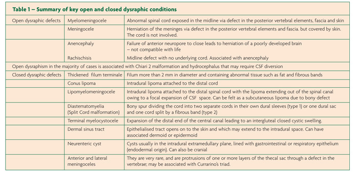
Spinal dysraphism is an umbrella term that describes any anomaly of the spinal cord, cauda equina or overlying tissues such as vertebrae, muscles and skin. The nervous system abnormality may or may not have associated mesenchymal or dermal changes.1,2,3 Spinal dysraphism is essentially an anatomical term describing a spectrum of lesions and associated pathology – tethered cord is the clinical manifestation of the anatomical abnormalities that constitute spinal dysraphism. Spinal dysraphism, also called spina bifida, can be subdivided into two groups: open/aperta and closed/occulta. Closed spinal dysraphism describes lesions with intact skin that are usually incidentally discovered on radiographic or physical exam. It is characterised by a disruption in the spinous processes and laminae (mesenchymal structures) without herniation of underlying abnormal or normal neural structures through the overlying skin. However open dysraphic lesions require emergent surgical repair to prevent infection, and are comprised of a broad spectrum of abnormalities. Table 1 summarises the key features of some open and closed dysraphic defects.
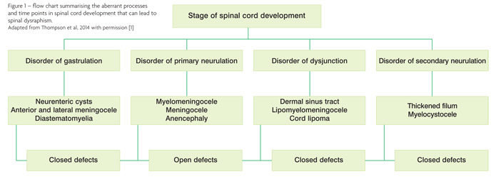
The spectrum of spinal dysraphism can also be classified according to the point in spinal cord embryological development that the abnormality occurs, which will be described in the following section on embryology of spinal dysraphism and in Figure 1.
Embryology
In order to understand the mechanism by which spinal dysraphism occurs, it is important to appreciate the normal embryology of spinal cord development. This is split into three key stages and at each stage abnormalities can arise that give rise to dysraphic lesions. Essentially closed spinal dysraphism is believed to be a problem of secondary neurulation (the third stage of spinal development) whilst open dysraphism is a problem earlier on, during primary neurulation.
Embryology of the normal spinal cord
Development of the normal spinal cord may be understood by viewing it as a three stage process: gastrulation, primary neurulation and secondary neurulation.
Gastrulation refers to the initial formation of a trilaminar plate that contains all three of the germ cell layers (ectoderm, mesoderm and endoderm) from which all future tissues will be derived. Ectoderm will give rise to the central nervous system and skin, endoderm to the viscera; and mesoderm to the musculo-skeletal system. During gastrulation, the primitive streak of the embryo gives way to development of the midline notochord. After gastrulation which lasts from days 16 to 18 primary neurulation occurs, followed by secondary neurulation.
Essentially primary neurulation will result in the formation of the brain and spinal cord whilst secondary neurulation refers to a separate process which forms the conus medullaris and cauda equina. Primary neurulation (days 18-28) involves the development of two ectodermal folds that fuse at the anterior and posterior neuropores as well as additional midline fusion points to form the brain (rostral end) and spinal cord (caudal end). This newly formed primitive central nervous system from the process of ectodermal fusion then undergoes “dysjunction” – physical separation from the rest of the ectoderm which then goes on to become skin. After dysjunction of neural tissue from the remainder of the ectoderm that is destined to be skin, the process of secondary neurulation (essentially caudal spinal development) can begin.
During secondary neurulation from days 28-48, a separate cell pool of pluripotent stem calls located at the very tail of the embryo, termed the “caudal cell mass” gives rise to the conus, cauda equina as well as parts of the genitourinary tract and the hindgut (the viscera being endodermal). The neural structures arising from the caudal cell mass join the distal spinal cord that was created by the prior process of primary neurulation. At this point, about eight weeks of gestation, spinal tissue extends down to the very bottom of the spinal column. The next stage is for the spinal cord to ascend rostrally in order for the conus to assume its usual position in the lumbar region. The basis of this process is by the bony vertebral column that houses the spinal cord growing disproportionately faster than the neural elements, resulting in the spinal cord being elevated proximally up the vertebral column. It is between the 8th and 18th week of gestation, that the vertebral column growth exceeds that of the spinal cord resulting in “caudal ascent” or migration of the cord to leave the thin filum terminale. By the 25th week of gestation, the growth subsides and, by two months of age the tip of the conus medullaris, the most caudal structure of the spinal cord, should be found between L1 and L2. A foetal post mortem MRI study showed that in 94.8% of cases, the conus had ascended to L3 by 40 weeks gestation.4 Ascension stops at three months.5,6 This process can be inhibited, however, in the presence of spinal dysraphism: the finding of the tip of the conus medullaris below the L1/2 junction can be considered pathological. A normally positioned conus does not exclude dysraphism.7
Occult dysraphic pathologies are believed to occur from failures of secondary neural tube closure and failures at the dysjunction stage.
Epidemiology
The incidence of dysraphism is approximately 1 per 500-1 per 1000 live births.8,9 The exact incidence of closed dysraphism is unknown but is significantly higher than that of open dysraphism, which is approximately 6 per 10,000;10 Open dysraphism is more common in female than in male children. In open dysraphism, women with a history of a previous pregnancy complicated by dysraphism carry a 3 to 5% risk of recurrent spinal dysraphism.11,12 The prevalence of all types of dysraphism that could be diagnosed at birth in the United States decreased almost a quarter between 1995-1996 and 2003-2004 following food fortification with folic acid.13
Both genetic and environmental factors have been implicated in the aetiology of spinal dysraphism. A number of genes have been investigated and no single causative gene has been identified, although genetic pathways in folate and 1-carbon metabolism are believed to be important.14 Environmental factors which have been demonstrated to be associated with high dysraphism risk are folate deficiency, use of some anti-epileptics, poor socio-economic status, maternal age greater than 40 or less than 19.15 Geographical variations in the prevalence of dysraphism have been reported in first and third world settings, although no firm conclusions regarding ethnicity being a risk factor have been reached.
The socioeconomic burden of dysraphism was investigated in a recent meta-analysis of 14 cost of illness studies which showed that key costs were hospital stay, long term complications of dysraphism throughout adulthood and caregiver costs.16
Known genetic syndromes associated with NTDs are trisomies 13 and 18 and Currarino triad17 and up to 10% of spina bifida cases are associated with chromosomal defects.18
Diagnosis and presentation
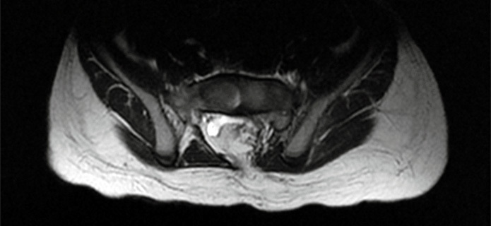
Open dysraphism is diagnosed by antenatal ultrasound and foetal MRI. The 2008 NICE guidelines on antenatal care recommend that when routine ultrasound screening is performed to detect spina bifida, it is not necessary to perform alpha-fetoprotein levels.19 The imaging is also useful to identify associated disorders such as hydrocephalus. Occult dysraphism is not usually identified antenatally. It is detected either when a midline cutaneous marker prompts further investigations or when the patient has an MRI for clinical suspicion of tethered cord or for other indications. Approximately 6% of closed dysraphism is associated with Chiari 1 malformation.20 It is therefore important to evaluate children with occult dysraphism for hydrocephalus and syringomyelia related to a Chiari 1.
Introduction to Tethered Cord Syndrome (TCS)
Tethered cord is essentially a clinical diagnosis. It refers to patients with dysraphic anomalies who have manifested clinical features of neurological, urological and orthopaedic pathology secondary to the dysraphism. The clinical heterogeneity of tethered cord syndrome makes the diagnosis a challenge. It may present at birth or be asymptomatic only to progress with growth and may present for the first time in adulthood.21 Finally, it may present as new or progressive signs in a patient with a known dysraphic state. The physical findings of occult spinal dysraphism do not necessarily correlate with the development of clinical tethered cord syndrome. Tethered cord syndrome is characterised by the progressive development of sensorimotor neurological dysfunction, sphincter and sexual dysfunction, and orthopaedic symptoms. These signs and symptoms often follow a period of growth, i.e. school age children between 5 and 15 years of age, and are the most common presentation.
Hypotheses for the pathophysiology of TCS
The terminology “tethered cord” refers to the fact that the cord or conus is low lying and was thought to have traction placed on it as the vertebral column grows whilst the nerve roots of the cauda equina are still adherent to the lumbosacral area and cannot ascend in parallel to the growing vertebral column. Nerve root stretch is theorised to result in vascular compromise with a decrease in blood flow and mitochondrial activity and metabolic derangement on the background of structurally abnormal neural tissue.22,23,24 Fibrous and fatty bands were identified in a histopathological and imaging study of dysraphism25 and these may cause mechanical “tethering”. These hypotheses have been debated in the literature, but the pathophysiology of the tethered cord syndrome remains unclear.
Cutaneous manifestations of the tethered cord syndrome
The main cutaneous stigmata of tethered cord are hairy patches, dermal sinuses, hemangiomas, subcutaneous lipomas, atretic meningoceles, abnormal dimples and skin tags.
Approximately half of clinically suspicious midline spinal cutaneous lesions are associated with dysraphic pathology.26 Approximately one third of patients with dysraphism show some cutaneous lesion.27 Skin manifestations are often single but less commonly can be multiple.
One lesion of spinal dysraphism is hypertrichosis, or the “hairy patch”. This lesion is described as a midline often V-shaped patch of excessive hair in the newborn, not a collection of sparse hair. Hairy patches associated with occult dysraphism are usually well defined and localised as opposed to hirsutism. Hairy patches can be associated with underlying split cord malformation.1
The finding of a lipoma, or a subcutaneous fat pad, is the most common marker that signifies underlying spinal dysraphism.28 In addition, 80-90% of spinal lipomas have an additional cutaneous lesion.29 The lipoma may be limited to the dermis, may extend into the intraspinal space via a vertebral defect, or may be limited to an intraspinal component, which would be a radiological finding only. A lipomyelomeningocele is a lesion which extends from the dermis to the intraspinal space, and should be ruled out prior to surgical resection of a midline spinal subcutaneous lipoma suspected to be purely superficial to the fascia.28
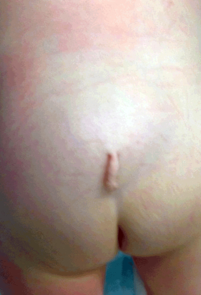
Dimples in the sacrococcygeal area often present as a finding on routine baby checks and pathological lesions are characterised by specific findings. Normal dimples are seen in 4% of infants and are characterised by their location at the tip of the coccyx, which is palpable through the dimple. The characteristics of a pathological dimple include those that are midline, located above the natal cleft and discharging. Dimples that are deeper or larger than normal also merit investigation and should be evaluated as markers of a dermal sinus tract. Dermal sinus tracts are epithelial lined conduits between the skin and underlying planes from superficial fascia down as far as the intradural space. Dermal sinuses are primarily of the midline lumbosacral spine (90%), although cases exist of thoracic and cervical dermal sinus tracts30 and tracts off the midline.31,32 They are believed to occur from failed separation between cutaneous and neural ectoderms – ie a failure of dysjunction.17 Dermal sinus tracts may be associated with dermoids and less commonly with epidermoids and rarely teratomas.17 Undetected, dermal sinuses can lead to meningitis and progressive neurological deficits. All children with recurrent meningitis should be assessed for a dermal sinus tract.
Midline lumbosacral skin discolouration (red or brown) or frank purple vascular malformations that are most worrisome are hemangiomas, which can be large and may be associated with other markers of dysraphism, such as dimples or hairy patches. Raised pigmented vascular lesions are usually associated with dysraphism.
Finally, the finding of a tail or pseudo-tail near the coccyx, or a midline raised skin tag anywhere along the spine, has a strong correlation with occult spinal dysraphism. A true tail is extremely rare and composed of fatty tissue, vasculature, muscle, and nerve fibres. All of these lesions are associated with occult spinal dysraphism and should prompt further evaluation.33
Unfortunately,not all occult dysraphic states are associated with a cutaneous marker, and those without cutaneous markers may present later when other clinical manifestations of dysraphism have developed.
Neurological, orthopaedic, bladder and bowel manifestations of TCS
Sensorimotor dysfunction can be from a combination of upper motor neuron (UMN) and lower motor neuron (LMN) dysfunction in the case of a conus lesion and pure LMN signs for lesions below the conus. This may present as lower extremity weakness and changes in sensation. Parents may report a delay in walking or changes in gait with the development of ataxia (worsening with fatigue or exercise), development of toe walking and regression of motor milestones. In addition, there may be contractures, alterations in tone, and muscle atrophy. The child may complain of back pain or radicular pain. Examination of the back may reveal scoliosis or abnormal lumbar lordosis in addition to the cutaneous markers described above.
Orthopaedic signs are essentially progressive deformities. They affect the lower limbs and/or the spine. Lower limb deformities are the result from imbalance between opposing muscle groups. These include pes cavus, varus, valgus and equinus deformities, tight Achilles tendons, clubbing, talipes, toe clawing, hammertoe, and leg length discrepancy. Spinal deformities can include scoliosis and sacral dysgenesis. These can be combined with deformity affecting individual vertebrae (segmental spinal dysgenesis) such as hemivertebrae, bifid vertebrae and laminar defects. Complications of orthopaedic lesions include low bone density,34 respiratory dysfunction and spasticity. TCS may be demonstrated through a sudden deterioration after straightening or correction in scoliosis is undertaken before untethering. Sometimes, the orthopaedic complaints may be addressed surgically without suspecting the underlying neurological problems.
TCS may also present with urological manifestations. These can affect the structure and function of the lower and upper urinary tracts. The most common urological problems are neurogenic bladder with associated vesicuoureteric reflux, overflow, recurrent urinary tract infections, stones and changes in continence. Presentation can be either with failure to attain continence or new onset incontinence. It may be subtle, with incomplete voiding, urinary frequency, stress incontinence and nocturnal enuresis.35 Up to 25% of children with occult dysraphism will have uncoordinated detrusor and sphincter activity, that can lead to permanent renal damage via high bladder pressures and recurrent urinary tract infections (UTIs).36 Sphincter function should be investigated early to avoid these complications.36 Other urological problems associated with spinal dysraphism include cryptorchidism, renal agenesis, horseshoe kidney and less commonly cloacal and bladder exstrophy.37
Bowel dysfunction without bladder involvement is rare in dysraphism.38 It may present with constipation or fecal incontinence. Rarely, imperforate anus may be present. Low and high anorectal malformations are associated with spinal dysraphism but are not usually directly visible without imaging.39,40 The finding of any of these signs and symptoms should prompt further evaluation into the possibility of spinal dysraphism as the etiology. Sometimes, urogenital, hindgut and dysraphic spinal pathologies can constitute one of the genetic syndromes, such as VACTERL (vertebral defects, anal atresia, cardiac defects, tracheo-esophageal fistula, renal anomalies, and limb abnormalities), OEIS (omphalocele, exstrophy of the cloaca, imperforate anus, and spinal defects) and Currarino’s triad (anorectal malformation, a sacral bony defect and a presacral mass).
Although it is well documented that sexual dysfunction can be a consequence of tethered cord and a complication of any treatment for it, the psychosexual effect of tethered cord is a poorly explored area, although from what evidence is available, only half these patients are satisfied with their sex life, mainly inhibited through incontinence and poor self image.41
Diagnosing tethered cord syndrome
The diagnosis of occult spinal dysraphism can be a challenging one to make, as the differential diagnosis is wide. The benefit unique to GPs is that of familiarity with the medical history of the child and family over time. Work up of TCS includes a thorough physical examination and neuroimaging. The physical examination is centred on cutaneous findings along with neurological, orthopaedic and sphincter features of cord tethering.
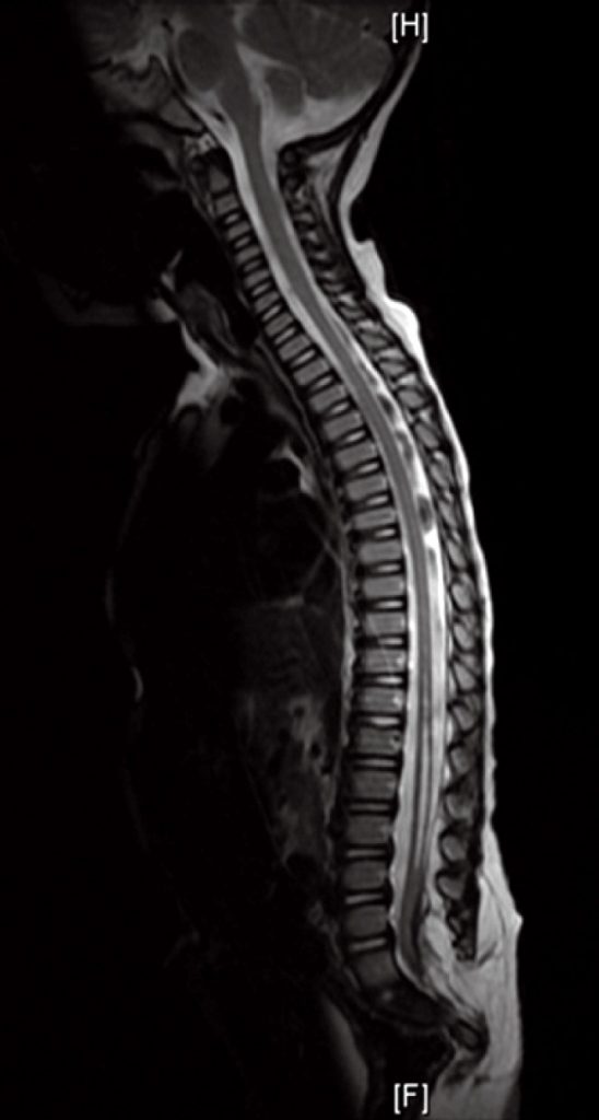
In addition to physical examination, concerns regarding this possibility should be evaluated with radiological studies. A spine x-ray can reveal bony abnormalities associated with a spinal dysraphism. However, it will not reveal any cord anatomy and causes unnecessary radiation exposure. For this reason, x-rays are obsolete now in the assessment of occult dysraphism. Ultrasonographic exam before the age of three months has been useful to examine the level of the conus and identify obvious dysraphic lesions. In addition, ultrasonography provides real time data and pulsations of the cord can be documented.42 But Magnetic Resonance Imaging is considered to be the “gold standard” in the investigation of spinal dysraphism. Absence of an abnormality on plain radiograph or with ultrasound evaluation in the face of a cutaneous marker or clinical findings should prompt further evaluation. If evidence of spinal dysraphism is found, a whole cranio-spinal MRI should be performed to identify any associated Chiari malformation, syringomyelia or hydrocephalus.
Options for managing TCS
Asymptomatic occult dysraphism is typically managed with regular outpatient follow up and active monitoring for orthopaedic, neurological and bladder/bowel changes that may suggest development of the tethered cord syndrome. The management of patients with occult dysraphism and clinical evidence of tethered cord syndrome is controversial and there is no level 1 evidence favouring a particular surgical approach or timing of surgery. The vast majority of surgeons would operate if the patient develops a new deficit related to tethered cord. Surgery is clearly indicated for those lesions placing the child in immediate danger, such as dermal sinus tracts associated with meningitis. The risks of untethering surgery are low when performed by an experienced Paediatric Neurosurgeon in a neurosurgical centre accustomed to this type of surgery. It is for these reasons that patients with radiological features of dysraphism will almost universally be referred for consideration of surgery. The case for prophylactic surgery is debated in the literature and is not undertaken in most centres on intact patients; although there is some evidence that surgery early in the course of clinical presentation, radical resections and surgery in children under the age of two years may be associated with favourable neurological outcome.43,44
In patients who undergo surgical management, the primary goal of the repair is to untether the cord and preserve or improve function. The secondary goal is the repair of the other associated anomalies available within the surgical exposure, such as lipomas and sinus tracts that currently or could in future contribute to tethering of the cord. This procedure may not extend to total resection of these anomalies if they are not responsible for the traction and tethering on the cord.
In surgery, patients are positioned prone and a midline incision is made centred over the area of pathology. The surgical approach tracks the abnormality from the skin, when present, through potentially abnormal subcutaneous tissues, fascia, bifid structures, and into the spinal cord or the filum terminale anomaly. The remainder of the operation focuses on the anatomical untethering of the attachments between the spinal cord and surrounding structures. Once the spinal cord is untethered, dural closure and closure of the overlying tissues is performed. The procedure is usually performed in a latex free environment to avoid sensitisation.
Spinal cord untethering is routinely performed under intraoperative neurophysiological motor tract and nerve root monitoring to allow stimulation of neurologically active structures and differentiation between adhesions that are functional from those that are not.45 Sensory potentials are of limited value.46 As complete a resection of the dysraphic anomaly causing TCS as possible should be performed to avoid retethering. The complication rate is quite low with experienced Paediatric Neurosurgeons. In a series of 238 cord lipoma resections, the incidence of neurological and urological complications was 4.2% and that of CSF leaks was 2.5%.47 In complex cases, only partial untethering may be possible.47
Active monitoring of conservatively managed and post operative patients is best performed by a multidisciplinary team composed of a Paediatrician, Neurosurgeon, Urologist, Rehabilitation Physician, Orthopaedic Surgeon and Physiotherapist. Monitoring consists of serial neurologic exams, imaging if new clinical features develop, orthopaedic follow up, and evaluation of bladder function. Serial urological exams with urodynamic evaluation provides an objective tool for the monitoring of abnormalities.
The role of the multidisciplinary team in the long term care of TCS patients
Issues presented to the care team in the immediate post-operative period are primarily those of wound integrity and hygiene. In children, especially those with low-lying lesions, wound infection becomes a significant risk. There is no evidence supporting prophylactic post operative antibiotics.
Another serious risk is that of wound breakdown and/or cerebrospinal fluid (CSF) leakage. Children may be placed on flat bed rest for 48 hours or more postoperatively to reduce the CSF pressure in the lumbar canal. The duration for maintaining a flat positioning is controversial. A longer period is often necessary after a tenuous dural repair. Open CSF leak out of the wound should be addressed with some urgency for fear of infection leading to meningitis. The CSF leak may however be contained under the skin in a pseudomeningocele. In this scenario, symptoms of a low-pressure headache may be present. External pressure dressings to obliterate subcutaneous dead space, CSF diversion or operative wound revision may need to be performed.
Despite surgical repair, a child with occult spinal dysraphism and/or tethered cord syndrome needs to be on close follow up throughout his or her life. The success rate of untethering is dependent on the cause of tethering, with more complex aetiologies making recurrence more likely.48 Most patients, after surgery, improve or have symptoms that stabilise. On the other hand, some (0-5%) may deteriorate. Deterioration is, as with the initial diagnosis of tethered cord syndrome, a clinical diagnosis49 and revision surgery focuses on identification and division of adhesions followed by a tight dural closure or duraplasty.48,49 Endoscopic division of adhesions may be performed by an experienced Paediatric Neurosurgeon.50 The MDT role is that of coordinating care as the child grows into adulthood. Children should be evaluated for signs of retethering, which has an associated lifetime risk of approximately 10%, higher for those in the subset with more complicated anatomical repairs.
Tethered cord patients are a heterogenous group of individuals in terms of their biopsychosocial care needs.51 Patients with associated hydrocephalus and a CSF shunt in situ are likely to have a greater biopsychosocial burden than patients without a shunt.52 This is due to any cognitive impairment from cranial pathology and also from shunt related complications. Long term musculoskeletal consequences of tethered cord include joint contractures, skin ulceration, spasticity, muscle wasting, chronic pain and gait disturbance. These translate into a variable degree of physical impairment and functional disability. Management of these requires input from Physiotherapists and Occupational Therapists. Close liaison between therapists, caregivers and educational institutions can be used to maximise the child’s participation in school activities. Care needs associated with a neurogenic bladder include training in home catheterisation, voiding techniques and regular follow up to monitor renal function. A number of non surgical and surgical strategies exist for the neurogenic bladder and bowel including enemas, stomas and bladder suspension.36 Urology specialist nurses and continence nurses can teach catheterisation, advise on hygiene and work with patients and their families to minimise the complications and social stigma associated with continence difficulties. Women with tethered cord need specialised care during pregnancy.53 In addition to the physical care requirements of the spina bifida patient, there are significant psychological and social aspects affecting the families of patients. A meta-analysis of 15 studies addressing parental psychological adjustment to spina bifida highlighted awareness of maternal psychological stress in particular.54 The involvement of Pain Physicians and Psychologists should be considered.55 A family centred rehabilitation model can address the needs of the family unit as well as the individual,56 but independently addressing the psychological care of the family, so it does not become entirely focused on the progress of the child.57
Throughout the child’s life, his/her normal development must be emphasised. This can truly be a challenge to the family faced with chronic health care issues. Stress should be placed on emphasising the normalcy of the child, particularly the normal cognitive development (in cases without co-existent cranial pathology that causes cognitive impairment), the need for normal treatment in relation to other children in the household, and the awareness of the family to the “vulnerable child” trap. Despite our inherent desire to protect the child from unnecessary difficulties or challenges in life, we may unfortunately contribute further to the handicap by doing so.
Conclusion
The finding of a midline spinal dysraphic defect in a child is often subtle and can be missed. Cutaneous lesions can, however, represent a marker of an occult spinal dysraphic state and therefore suspicious lesions need further evaluation. The necessity of evaluation is due to the possibility of a spinal dysraphic state that can lead to the clinical entity of tethered cord syndrome. This diagnosis, its subsequent evaluation and management requires a multidisciplinary team, headed by the child’s primary caregiver. Management options continue to be debated as not all low-lying spinal cords progress to tethered cord syndrome. Regardless, the role of the General Practitioner and the Paediatrician in the initial evaluation and communication to families of these possibilities, and his/her awareness to the impact of these diagnoses is critical in the optimisation of the child’s physical and developmental health.
References
- Thompson D. Spinal dysraphic anomalies; classification, presentation and management. Paediatrics and Child Health 2014.
- Thompson D. Spinal dysraphic anomalies; classification, presentation and management. Paediatrics and Child Health. Volume 20, Issue 9, Pages 397–403, September 2010.
- Frim D. Spinaldysraphism. Chapter: http://www.landesbioscience.com/vademecum/Frim_9781570597503.pdf ISBN 978-1-57059-643-8
- Arthurs OJ, Thayyil S, Wade A et al. Magnetic Resonance Imaging Autopsy Study Collaborative Group. Normal ascent of the conus medullaris: a post-mortem foetal MRI study. J Matern Fetal Neonatal Med. 2013 May;26(7):697-702.
- Hawass ND, el-Badawi MG, Fatani JA et al. Myelographic study of the spinal cord ascent during fetal development. AJNR Am J Neuroradiol. 1987 Jul-Aug;8(4):691-5
- Yamada S, Perot PL Jr, Ducker TB et al. Myelotomy for control of mass spasms in paraplegia. J Neurosurg. 1976 Dec;45(6):683-91.
- Tubbs RS, Oakes WJ. Can the conus medullaris in normal position be tethered? Neurol Res 2004;26:727–731.
- Mitchell LE. Epidemiology of neural tube defects. Am J Med Genet C Semin Med Genet. 2005 May 15;135C(1):88-94. Review.
- Parker SE1, Mai CT, Canfield MA et al. Updated National Birth Prevalence estimates for selected birth defects in the United States, 2004-2006. Birth Defects Res A Clin Mol Teratol. 2010 Dec;88(12):1008-16.
- Boyd PA, Tonks AM, Rankin J et al. Monitoring the prenatal detection of structural fetal congenital anomalies in England and Wales: register-based study. Journal of Medical Screening
2011;18(1):2. - Blatter BM, Lafeber AB, Peters PW et al. Heterogeneity of spina bifida. Teratology 1997Apr;55(4):224-30.
- Cowchock S, Aunbender E, Precott G et al. The recurrence risk for neural tube defects in the United States: A collaborative study. Am J Med Genet 1980;5:309-314.
- Boulet SL, Yang Q, Mai C et al. Trends in the postfortification prevalence of spina bifida and anencephaly in the United States. National Birth Defects Prevention Network. Birth Defects Res A Clin Mol Teratol. 2008 Jul;82(7):527-32.
- Beaudin AE, Stover PJ. Insights into metabolic mechanisms underlying folate-responsive neural tube defects: a minireview. Birth Defects Res A Clin Mol Teratol. 2009 Apr;85(4):274-84.
- Vieira AR, Castillo Taucher S. Maternal age and neural tube defects: evidence for a greater effect in spina bifida than in anencephaly. Rev Med Chil. 2005 Jan;133(1):62-70.
- Yi Y, Lindemann M, Colligs A et al. Economic burden of neural tube defects and impact of prevention with folic acid: a literature review. Eur J Pediatr. 2011 Nov;170(11):1391-400.
- Özek, M. Memet, Cinalli, Giuseppe, Maixner, Wirginia (Eds.). Spina Bifida. Management and Outcome. 2008. ISBN 978-88-470-0651-5.
- Sepulveda W, Corral E, Ayala C et al. Chromosomal abnormalities in fetuses with open neural tube defects; prenatal identification with ultrasound. Ultrasound Obstet Gynecol 2004;23:352-6.
- Antenatal care. Routine care for the healthy pregnant woman http://www.nice.org.uk/nicemedia/pdf/CG062NICEguideline.pdf
- Valentini LG, Selvaggio G, Visintini S et al. Tethered cord: natural history, surgical outcome and risk for Chiari malformation 1 (CM1): a review of 110 detethering. Neurol Sci. 2011 Dec;32 Suppl 3:S353-6.
- Pang D, Wilberger JE Jr. Spinal cord injury without radiographic abnormalities in children. J Neurosurg. 1982 Jul;57(1):114-29.
- Filippidis AS, Kalani MY, Theodore N et al. Spinal cord traction, vascular compromise, hypoxia, and metabolic derangements in the pathophysiology of tethered cord syndrome. Neurosurg Focus. 2010 Jul;29(1):E9.
- Stetler WR Jr, Park P, Sullivan S. Pathophysiology of adult tethered cord syndrome: review of the literature. Neurosurg Focus. 2010 Jul;29(1):E2.
- Yamada S, Iacono RP, Andrade T et al. Pathophysiology of tethered cord syndrome. Neurosurg Clin N Am. 1995 Apr;6(2):311-23. Review
- Thompson EM, Strong MJ, Warren G et al. Clinical significance of imaging and histological characteristics of filum terminale in tethered cord syndrome. J Neurosurg Pediatr.
2014 Mar;13(3):255-9. - Tavafoghi V, Ghandchi A, Hambrick GW Jr et al. Cutaneous signs of spinal dysraphism. Report of a patient with a tail-like lipoma and review of 200 cases in the literature. Arch Dermatol. 1978 Apr;114(4):573-7.
- Yamada S. Tethered Cord Syndrome in Children and Adults. 2nd edition. ISBN (Americas): 9781604062410
- Dias MS, Li V. Paediatric neurosurgical disease. Pediatr Clin North Am, 1998;45(6):1539-78.
- Gorey MT, Naidich TP, McLone DG, Double discontinuous lipomyelomeningocele: CT findings. J Comput Assist Tomogr, 1985;9(3):584-91.
- Ackerman LL, Menezes AH, Follett KA. Cervical and thoracic dermal sinus tracts. A case series and review of the literature. Pediatr Neurosurg. 2002 Sep;37(3):137-47.
- Ikwueke I, Bandara S, Fishman SJ et al. Congenital dermal sinus tract in the lateral buttock: unusual presentation of a typically midline lesion. J Pediatr Surg. 2008 Jun;43(6):1200-2.
- Steinbok P. Dysraphic lesions of the cervical spinal cord. Neurosurg Clin N Am. 1995 Apr;6(2):367-76.
- Belzberg AJ1, Myles ST, Trevenen CL. The human tail and spinal dysraphism. J Pediatr Surg. 1991 Oct;26(10):1243-5.
- Szalay EA, Cheema A. Children with spina bifida are at risk for low bone density. Clin Orthop Relat Res. 2011 May;469(5):1253-7.
- Shin SH, Im YJ, Lee MJ et al. Spina bifida occulta: not to be overlooked in children with nocturnal enuresis. Int J Urol. 2013 Aug;20(8):831-5.
- de Jong TP, Chrzan R, Klijn AJ et al. Treatment of the neurogenic bladder in spina bifida. Pediatr Nephrol. 2008 Jun;23(6):889-96.
- Netto JM, Bastos AN, Figueiredo AA et al. Spinal dysraphism: a neurosurgical review for the urologist. Rev Urol. 2009 Spring;11(2):71-81.
- Thompson D. Hairy backs, tails and dimples. Current Paediatrics 2000;10:177-83.
- Rivosecchi M, Lucchetti MC, Zaccara A et al. Spinal dysraphism detected by magnetic resonance imaging in patients with anorectal anomalies: incidence and clinical significance. J Pediatr Surg. 1995 Mar;30(3):488-90.
- Warf BC, Scott RM, Barnes PD et al. Tethered spinal cord in patients with anorectal and urogenital malformations. Pediatr Neurosurg. 1993;19(1):25-30.
- Verhoef M, Barf HA, Vroege JA et al. Sex education, relationships, and sexuality in young adults with spina bifida. Arch Phys Med Rehabil. 2005 May;86(5):979-87.
- Lowe LH, Johanek AJ, Moore CW. Sonography of the neonatal spine: part 2, Spinal disorders. AJR Am J Roentgenol. 2007 Mar;188(3):739-44.
- Pang D, Zovickian J, Oviedo A.Long-term outcome of total and near-total resection of spinal cord lipomas and radical reconstruction of the neural placode, part II: outcome analysis and preoperative profiling. Neurosurgery. 2010 Feb;66(2):253-72; discussion 272-3.
- Tseng JH1, Kuo MF, Kwang Tu Y et al. Outcome of untethering for symptomatic spina bifida occulta with lumbosacral spinal cord tethering in 31 patients: analysis of preoperative prognostic factors. Spine J. 2008 Jul-Aug;8(4):630-8. doi: 10.1016/j.spinee.2005.11.005. Epub 2006 Jul 11.
- Hoving EW, Haitsma E, Oude Ophuis CM. The value of intraoperative neurophysiological monitoring in tethered cord surgery. Childs Nerv Syst. 2011 Sep;27(9):1445-52.
- Li V, Albright AL, Sclabassi R et al. The role of somatosensory evoked potentials in the evaluation of spinal cord retethering. Pediatr Neurosurg. 1996;24(3):126-33.
- Pang D, Zovickian J, Oviedo A. Long-term outcome of total and near-total resection of spinal cord lipomas and radical reconstruction of the neural placode: part I-surgical technique. Neurosurgery. 2009 Sep;65(3):511-28; discussion 528-9.
- Samuels R, McGirt MJ, Attenello FJ et al. Incidence of symptomatic retethering after surgical management of pediatric tethered cord syndrome with or without duraplasty. Childs Nerv Syst. 2009 Sep;25(9):1085-9.
- Shih P1, Halpin RJ, Ganju A et al. Management of recurrent adult tethered cord syndrome. Neurosurg Focus. 2010 Jul;29(1):E5.
- Di X. Endoscopic spinal tethered cord release: operative technique. Childs Nerv Syst. 2009 May;25(5):577-81.
- Fletcher JM and Brei TJ. Introduction: Spina bifida–a multidisciplinary perspective. Dev Disabil Res Rev. 2010;16(1):1-5.
- Ramachandra P, Palazzi KL, Skalsky AJ et al. Shunted hydrocephalus has a significant impact on quality of life in children with spina bifida. PM R. 2013 Oct;5(10):825-31.
- Stansfield C. Maternity care of a woman with spina bifida. Pract Midwife. 2012 Jun;15(6):34-6.
- Vermaes IP, Janssens JM, Bosman AM et al. Parents’ psychological adjustment in families of children with spina bifida: a meta-analysis. BMC Pediatr. 2005 Aug 25;5:32.
- Engel JM, Wilson S, Tran ST et al. Pain catastrophizing in youths with physical disabilities and chronic pain. J Pediatr Psychol. 2013 Mar;38(2):192-201.
- Piškur B1, Beurskens AJ, Jongmans MJ et al. Parents’ actions, challenges, and needs while enabling participation of children with a physical disability: a scoping review. BMC Pediatr. 2012 Nov 8;12:177.
- Ulus Y, Tander B, Akyol Y et al. Functional disability of children with spina bifida: its impact on parents’ psychological status and family functioning. Dev Neurorehabil. 2012;15(5):322-8.


