Abstract
Arabic texts of the ninth century described loss of sight as one form of ocular paralysis. Some early descriptions of amaurosis in retrospect probably describe optic neuropathy but its nature and defining physical signs arose from Helmholtz’s ophthalmoscope in 1845. In 1864 von Gräfe and later Thomas Buzzard and Clifford Allbutt gave detailed accounts, but the most important description was the 1884 work of the ophthalmologist Edward Nettleship, which is here recounted.

Optic neuritis, often named retrobulbar neuritis or optic papillitis, is one of the commonest symptoms of multiple sclerosis (MS). At some stage it affects over 50% of MS patients. An early description in 1864 was by Albrecht von Gräfe [Graefe] (1828-1870) [1]. Modern techniques made diagnosis easier and more precise, but the early descriptions and much-argued concepts are seldom discussed. A thinning of the serial ganglion cell layer and inner plexiform layer is the pathogenesis of acute optic neuritis.
Ancient references to optic nerve dysfunction as a mechanism for loss of vision are found in Arabic texts of the ninth century [2]. Possibly the first textbook of ophthalmology was written by Hunayn ibn Ishaq, (808-873) a Nestorian Christian and chief physician to the Caliph al-Mutawakkil. Like Galen, he believed that the optic nerve was hollow to transmit psychic pneuma* that flowed from the brain; the lens was the organ of vision. This he deduced by shutting one eye, whereupon the pupil of the other became enlarged to allow the escape of diverted pneuma. When the closed eye was opened, the enlarged pupil contracted to normal size [3]. He described three different forms of ocular paralysis: those involving sight alone, those involving eye movements alone, and those involving both; but he failed to separate optic neuritis from other eye diseases:
The vision has ceased or diminished without our finding any change in the pupil and there is heaviness in the head and particularly its deep part and the parts surrounding the orbit. We know that the affection is caused by abundant moisture, which has run to the optic nerve…
Even before von Gräfe, in December 1822, Sir Augustus D’Este, grandson of King George III., when he was 28 years old, suffered what in retrospect was an attack of retrobulbar neuritis, though its nature was not realised at the time. In successive years, he noted progressive weakness, numbness, difficulty in walking, painful spasms and depression – all typical of MS [4,5]. Although he was aged 54 when he died, no formal diagnosis was made, but ‘the meticulous notes in his diary justify a posthumous diagnosis [5]’.
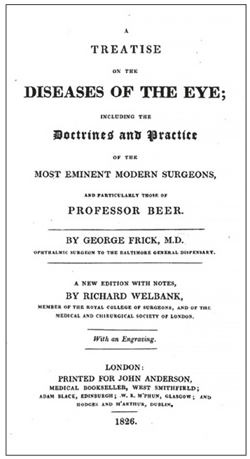
In 1823, George Frick (1793-1870) [4] in the first American textbook (Figure 1) of ophthalmology (1823) had described varieties of amaurosis that included but did not demarcate optic neuritis:
The terms amaurosis, gutta serena, suffusion nigra, &c are applied to a species of blindness which is produced by some immediate affection of the optic nerve or its expansion into the retina … Amaurosis may take place suddenly or slowly and be transient, permanent or intermittent. (Pp.138-141. 1826 edition)
But before the ophthalmoscope, Frick was unable clearly to distinguish optic neuritis from uveitis, glaucoma, orbital tumours, and other eye and systemic disorders. Shrewdly, he had noted severe pain in the orbit before visual loss and abnormal pupillary responses to light. He related:
Amaurosis from whatever cause … is generally characterized by a very dilated pupil which is not affected by any degree of light which is made to fall upon the retina … [the patient] is obliged to turn his head to render them [objects] distinct. (p. 142)
The invention of the ophthalmoscope by Hermann von Helmholtz (1821–1894) in 1845 [5,6] allowed optic neuritis to be separated from many other ocular disorders. So valuable was the ophthalmoscope that by 1871, Thomas Clifford Allbutt [8] (1836-1925) protested:
The number of physicians who are working with the ophthalmoscope in England may, I believe, be counted upon the fingers of one hand.
And in this seminal text, he recognised the salient features of optic neuritis and ‘atrophic amaurosis’ and the frequent confusion with ischaemic optic neuropathy [7].
In 1860 von Graefe ((1828–1870) [8], and more meticulously, Edward Nettleship (1845-1913) in 1884 described its principal features [9]. Nettleship acknowledged that both Leber and Jonathan Hutchinson had previously described cases of optic neuritis. However, Leber included instances of tobacco amblyopia and Hutchinson included several other pathologies. Nettleship’s comprehensive account in 1884 emphasised pain on eye movement, abnormal disk appearances and he stressed the impaired colour vision. Eleven of his 16 patients had a central scotoma. He accurately characterised its features:
Failure of sight limited to one eye, often accompanied by neuralgic pain about the temple and orbit and by pain in moving the eye; many recover but permanent damage and even total blindness may ensue; there is at first little, sometimes no, ophthalmoscopic change, but the disc often becomes more or less atrophic in a few weeks… The defect in vision is often described at first as a ‘‘gauze’’ or a ‘‘yellow mist’’ or a ‘‘dark patch’’ or a ‘‘spot’’ which covers the object looked at and gives an unnatural colour, the hand looking, for example, as if covered by a brown glove.
Although he identified all the salient features of optic neuritis, he did not mention a relationship to other relapsing and remitting neurologic symptoms, characteristic of MS. In the 19th century, optic neuritis was often used as a descriptive term for papilloedema. Its most common cause was widely said to be a brain tumour. It was also recognised as a discrete disease of the optic nerves [9], though its aetiology was often uncertain.

After Nettleship’s seminal description, Thomas Buzzard (1831-1919)† (Figure 2) in 1893 reported five patients with a history of disseminated sclerosis, who had episodes of visual failure with recovery consistent with optic neuritis [10]. He was one of the first to recognise optic atrophy as a feature of disseminated (multiple) sclerosis.
He credited Charcot with the first description of Sclérose en plaques and for noting amblyopia as a frequent symptom [11]. Parinaud [12] had noted the impairment of colour vision in optic neuritis, and later James Adie re-emphasised the importance of a central or paracentral scotoma [13], which he regarded as essential for diagnosis.
Wilhelm Uhthoff (1853–1927) [14], a Privatdocent für Augenheilkunde in Berlin, in 1890 described characteristic, transient blurring of central vision on exercise, rise in temperature, and fatigue in disseminated sclerosis, known as Uhthoff’s sign.
Neuromyelitis optica (Devic’s disease)
In 1870, (Sir) Thomas Clifford Allbutt, then Physician to the General Infirmary at Leeds, first reported the association between myelitis and an optic nerve disorder [15]. He briefly described a case of myelitis followed by optic neuritis approximately three months later. Earlier, EO Hocken had reported a patient with spinal cord inflammation and amaurosis in 1841 [16]. CM Durrant described a probable case in 1850 [17]. Wilhelm Erb in 1879 described a man who developed recurrent optic neuritis succeeded by subacute myelitis [18]. Dreschfeld in 1882 described the first pathologically examined case [19] and showed inflammation in both the spinal cord and optic nerves; the brain was normal. In 1894, Eugène Devic (158-1930) [20] presented his case at the First Congress of Internal Medicine in Lyon, and with Gault summarised 16 patients with loss of vision, who within weeks developed spastic paresis. Devic’s telling question, ‘Why such a peculiar localisation?’ remains unanswered. Neuromyelitis optica (NMO) has well-known similarities to multiple sclerosis, but despite the ending of NMO-IgG (Aquaporin 4) in about two-thirds of cases of NMO, whether it is an Aquaporin 4 + NMO autoimmune astro-cytopathy, a disease sui generis, or an MS variant remains arguable [21].
In typical optic neuritis, visual function improves spontaneously over four to six weeks, and within 12 months 93% have acuity of at least 20/40. High-dose corticosteroids may hasten recovery, but have little effect on long-term visual outcome. The cumulative probability of developing MS by 15 years is 50%. White matter plaques on the first magnetic resonance image increase that risk to 72%.
Edward Nettleship (1845-1913)
Of the many who contributed to the descriptions of optic neuritis it is Nettleship whose comprehensive writings first clearly delineated the disorder. Born on 3rd March 1845 in Kettering, Northamptonshire, he attended Kettering grammar school. Intending at first to become a farmer, he entered King’s College, London, and the Royal Veterinary College, and was admitted a Licentiate of the Society of Apothecaries and received the diploma of the Royal College of Veterinary Surgeons in 1867. He was appointed Professor of Veterinary Surgery at the Royal Agricultural College but a year later returned to the London Hospital, as dresser and assistant to (Sir) Jonathan Hutchinson [22]. Nettleship became his firm friend, and most distinguished pupil. He qualified in surgery (FRCS), from the London Hospital and the Blackfriars Hospital for Skin Diseases. To specialise in ophthalmology, in 1868, he studied at Moorfields Eye Hospital under Hutchinson and Waren‡ Tay. He was appointed Curator of the Museum and Librarian.
On 22 January 1869 he married Elizabeth Endacott Whiteway from Devon; they had no children. At Moorfields he began his researches into eye diseases [23], but left for the post of medical superintendent at the Ophthalmic School at Bow (1873-4), working with impoverished children suffering from eye infections, He was then appointed ophthalmologist at the South London Ophthalmic Hospital, St Thomas’s Hospital (1878-95), and surgeon at Moorfields (1882-98). In 1888 he became Dean of the Medical School. He served and became President of the Ophthalmological Society, and advised the Board of Trade on Sight Tests for the Mercantile Marine.
Nettleship acquired a considerable reputation as an eye surgeon and teacher. Sir John Parsons described him as the most scientific teacher of his time. He removed a cataract from William Ewart Gladstone, and attended Queen Victoria for the same condition, but advised against surgery.

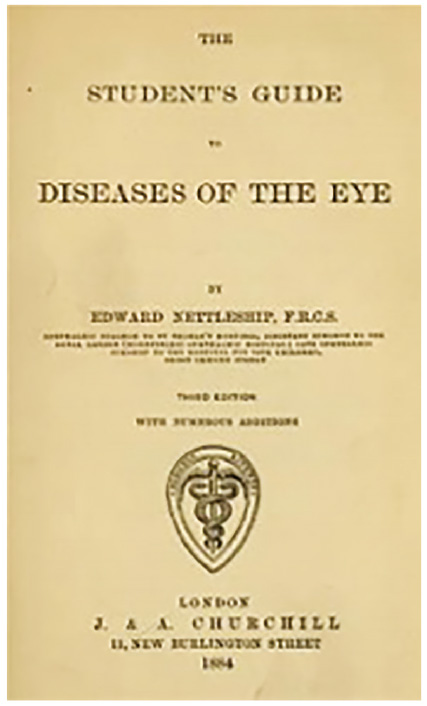
Whilst working at St Thomas’s and Moorfields Eye hospital, he wrote the definitive text: The Student’s Guide to Diseases of the Eye (Figure 3) that ran to five editions. His On Some Hereditary Diseases of the Eyes, 1909 was the standard monograph. His studies on hereditary eye diseases: albinism, retinitis pigmentosa, were executed mainly in ‘retirement’ in 1902 to Hindhead; there he concentrated on a pioneering series of papers on hereditary eye disease which was recognised in 1912 by the Fellowship of the Royal Society. Nettleship, a shy, reserved man, attracted many disciples to his clinic, who were keen to learn his methods and to submit to his somewhat severe discipline. But his patients and friends readily appreciated his underlying kindness and generosity. Prostatic surgery in 1911 led to distressing complications, accompanied by colonic carcinoma. Despite radiotherapy, he died at his home, in Hindhead, Surrey, on 30 October 1913. The Ophthalmological Society awards the Edward Nettleship Prize triennially; the first was to Nettleship himself in 1909 in recognition of his researches.
*Pneuma: metaphorical breath of life
† One of his sons was Sir Farquhar Buzzard, Bart, F.R.C.P, who followed in his footsteps as physician to the National Hospital, Queen Square
‡ Usually misspelt Warren
References
- von Gräfe ‘ Klin, Monatsbl f Augeuh, i, 49 cited in: Clinical lecture on a case of retro-bulbar abscess. Medicine, Surgery, And Their Allied Sciences. The New Sydenham Society. London. MDCCCLXIV. p. 267
- Volpe NJ. Optic Neuritis: Historical Aspects. Journal of Neuro-Ophthalmology 2001;21(4):302-9. https://doi.org/10.1097/00041327-200112000-00015
- Hunain ibn Is-hâq. The Book of the Ten Treatises on the Eye, Publisher: English translation and glossary by Max Meyerhof Government Press: Cairo, 1928.
- Frick G. A treatise on the diseases of the eye. Baltimore: Fielding Lucas; 1823.
- Helmholtz H. Beschreibung eines Augen-Spiegels. Berlin, Germany: A Förstner’sche Verlagsbuchhandlung; 1851. https://doi.org/10.1007/978-3-662-41295-4
- Pearce J.M.S. The Ophthalmoscope: Helmholtz’s Augenspiegel. Eur Neurol 2009;61:244-9.
https://doi.org/10.1159/000198418 - Allbutt TC, Allbutt T. On the ophthalmoscopic signs of spinal disease. Lancet 1870;l:76-8. https://doi.org/10.1016/S0140-6736(02)68218-2
- von Graefe A. Ueber complication von sehnervenentzundung mit gehirnkrankheiten. Archiv Ophthalmologic 1860;1:58-71.
- Nettleship E. On cases of retro-ocular neuritis. Trans Ophthal Soc UK 1884;4:186-226.
- Buzzard T. Atrophy of the optic nerve as a symptom of chronic disease of the central nervous system. Br Med J 1893;2:779-784.
- Charcot JM. Histologie de la sclérose en plaques. Gaz Hop (Paris) 1868;41:554, 557-8, 566.
- Parinaud H. Troubles oculaires de la sclerose en plaques. J Sante 1884;3:3-5.
- Adie W J. The aetiology and symptomatology of disseminated sclerosis. British Medical Journal 1932;2:997-1000. https://doi.org/10.1136/bmj.2.3752.997
- Uhthoff W. Untersuchungen uber die bei der multiplen herdskle- rose vorkommenden augenstorungen. Arch Pyschiat NervKranken 1890;21:55-116, 303-410. https://doi.org/10.1007/BF02162972
- Allbutt T. On the ophthalmoscopic signs of spinal disease. Lancet 1870;l:76-8. https://doi.org/10.1016/S0140-6736(02)68218-2
- Hocken E (1841) Illustrations of the pathology and treatment of the amauroses. Amaurosis from affections of the spinal cord or its membranes. London Medical Gazette 707. XXVIII. New Series. Vol. II. 1840-41:499-504. Cited by Jarius, S. & Wildemann, B. J Neurol (2014) 261: 400. doi:10.1007/s00415-013-7210-x
https://doi.org/10.1007/s00415-013-7210-x - Jarius S, Wildemann B. An early case of neuromyelitis optica (1850: Provincial Medical and Surgical Journal). Brit Med J 2012;345:e6430. https://doi.org/10.1136/bmj.e6430
- Erb W. Uber das Zusammenkommen von Neuritis optica und Myelitis subacute. Arch Psychiatr Nervenkr 1879;1:146-57. https://doi.org/10.1007/BF02224560
- Dreschfeld J. Pathological contributions on the course of the optic nerve bres in the brain. Brain 1882;4:543-51. https://doi.org/10.1093/brain/4.4.543
- Devic E. Myelite subaigue compliquee de neurite optique. Bull Med. 1894;8:1033-4.
- Weinshenker BG, Wingerchuk DM. The two faces of neuromyelitis optica. Neurology February 11, 2014 vol. 82 no. 6 466-7. https://doi.org/10.1212/WNL.0000000000000114
- Plarr’s Lives of the Fellows Online. Nettleship, Edward (1845 – 1913)
- Branford WA. ‘Nettleship, Edward (1845-1913)’, Oxford Dictionary of National Biography, Oxford University Press, 2004 [http://www.oxforddnb.com/view/article/35201, accessed 29 April 2017] https://doi.org/10.1093/ref:odnb/35201
- Bukhari W, Prain KM, Waters P, et al Incidence and prevalence of NMOSD in Australia and New Zealand. J Neurol Neurosurg Psychiatry Published Online First: 26 May 2017. doi: 10.1136/jnnp-2016-314839. https://doi.org/10.1136/jnnp-2016-314839