Introduction to the ACNR Stroke Series
In the second article in the ACNR stroke series, we are delighted to have an article by Alex Leff on recovery of language and vision, key challenges in stroke rehabilitation where new insights into mechanisms have developed rapidly in recent years, with the beginnings of evidence-based treatments. Alex has been a pioneer in these fields, especially in translating new understanding of reading problems after stroke into web-based solutions for both treatment delivery and data collection. In this clear and succinct summary, he outlines the latest advances and challenges for the future.
David Werring, Reader in Clinical Neurology, UCL Institute of Neurology, National Hospital for Neurology and Neurosurgery, Queen Square, WC1N 3BG.
Language
Normal language function is usually dependent on an intact left-hemisphere; however, the current neuroscientific evidence suggests that individual language functions are supported by a network of nodes and connections [1]. For instance, naming is subserved by an extensive network that is vulnerable to damage at many of its nodes which is why most aphasic patients suffer from anomia. Acquired language impairment (dysarthria or aphasia) is the second commonest major symptom caused by stroke, with an estimate prevalence of 250,000 in the UK.
Evidence base for speech therapy for aphasia
Despite occasional high profile papers that appear to show that speech and language therapy (SALT) is no better than non-specialist interventions [2], the evidence for the overall effectiveness of SALT is pretty clear, even Cochrane agrees: “We identified 39 trials involving 2518 randomised participants that were suitable for inclusion in this review. Overall, the review shows evidence from randomised trials to suggest there may be a benefit from speech and language therapy but there was insufficient evidence to indicate the best approach to delivering speech and language therapy [3].”
Bhogal’s meta-analysis treated the data in an interesting way, segregating positive from negative SALT studies and asking what the systematic differences between them were [4]. The answer was compelling: positive studies delivered ~100 hours of therapy, while patients in negative studies clocked-up only 45 hours of therapy. Unfortunately, the average out-patient NHS dose is 8-12 hours in total [5].
Future for SALT
Given what we know about the total dose required and the lack of SALT time, computer-based or web-based therapies are probably the most promising way to deliver enough dose of appropriate SALT to aphasic patients. There are many e-therapy devices or programmes available with over 50 software programs and over 40 apps: http://www.aphasiasoftwarefinder.org. Unfortunately, almost none have been subject to a clinical trial.
Quite a large range of drug therapies have been trialed in aphasia. Well-designed studies (placebo-controlled, double-blind) have been carried out that show significant improvements when L-dopa, memantine or donepezil are used. In studies where the drug was paired with behavioural therapy, the SALT effect sizes average at a fairly consistent 0.3 while the drug effects can be substantially higher than this but are patchy by comparison [6].
Another promising adjuvant is transcranial direct current stimulation (tDCS). In this technique a small current is passed through the brain using a battery and scalp electrodes. Because the current is continuous, there is no sensory stimulation to the scalp making this an easy technique to double-blind. It is not yet clear which regime to use (strength of current, exact placement of leads, activation of left, right or both hemispheres) [7] but if tDCS effects on language follow in the foot-steps of motor system research, then modest gains of about a third, compared with the effect of practice alone, can be expected.
Exactly when therapy should be delivered is still an open question. “As soon as possible” seems an obvious answer but the jury is still out on the effects of high-intensity speech therapy in the acute phase. It is important to remember that with survival rates increasing (current five-year survival is now at 82% in the UK [8]), the vast majority of aphasics are in the chronic phase, where NHS therapy resources are scant. Importantly, in terms of years post-stroke, there appears to be no upper limit beyond which patients cannot improve if they have access to targeted therapy [9].
Vision
The many interventions for hemianopia can be classified into three main groups: visual restitution, eye movement therapy (a strategy-based therapy) and visual aids. The difference between the last two categories is that strategy-based therapies transfer gains to function outside the periods of therapy, whereas aids improve function while being used but have no effect on behaviour once removed (wearing glasses improves your visual acuity, but not when you take them off).
Hemianopia is very common with stroke alone responsible for ~15,000 new cases per year in the UK. Unlike in the language or motor system where therapy induced gains can be seen years or even decades after stroke, visual field defects rarely improve six months after the onset [10]. Hemianopias that encroach on central vision (the majority) affect a whole host of activities of daily living [11].
Evidence base for hemianopia therapies
There are two main types of visual restitution therapies and each comes with its own patented hardware. One aims to improving high-acuity, conscious vision at the borders of the field defect, NovaVision VRT (visual restoration therapy); while the other, Neuro-Eye therapy, improves vision deep into the impaired field. Both rely on mass practice of detecting stimuli. There has been much controversy over whether NovaVision VRT really is effective, and, if so, by what mechanism. The main issue has been that any visual field gains have only been seen using the visual field test that is bundled with the therapy package. This is a problem as the visual field test relies on the same stimuli that are used in the training or therapy part of the programme. Obviously, if vision is really being restored, it shouldn’t matter what method is being used to test visual field gains. The prevailing view is that the training results in more efficient eye movements into the damaged field and not genuine field expansion. Patients themselves care much less about the mechanism of action than dueling scientists, and there is good evidence that the therapy does improve visual function.
Neuro-Eye therapy is less controversial as the stimuli (Gabor patches) are presented deep into the blind field and probably rely on sparse, non-V1 pathways by which visual stimuli can be processed but do not always reach conscious perception (the definition of blindsight). Practice effects are retinotopic, that is, performance gains are only seen in those parts of the visual field where stimuli were shown although this requires thousands to tens-of-thousands of trials. The question is: how useful is blindsight? It is certainly better than nothing and may help reduce patients colliding with objects, but there have been no studies addressing the impact of this training regime on activities of daily living.
The most convincing and consistent evidence for improving visual function comes from studies that retrain eye movements. This involves mass practice using stimuli that provoke a specific type of eye movement. Unlike the visual restitution therapies these have a clear carry-over effect onto untrained stimuli. There are over 10 published studies in this area and a recent Cochrane review of the more rigorous ones concluded that, “There is limited evidence which supports the use of compensatory scanning training for patients with visual field defects [12].” A further study published since then very nicely demonstrates the specificity of these techniques [13]. Using a randomised, cross-over design of reading therapy and visual exploration training, the results showed that the training-related improvements in reading and visual exploration were specific and task dependent; the combined effect size was 0.47 for visual search and 0.28 for reading. Interestingly, the cross-over therapy had no effect (positive or negative) on the other task; that is, when undergoing the second therapy block, gains made from the first block remained stable. While the specificity was no great surprise (although this was the first study to explicitly test this) it is good news that there was no interference.
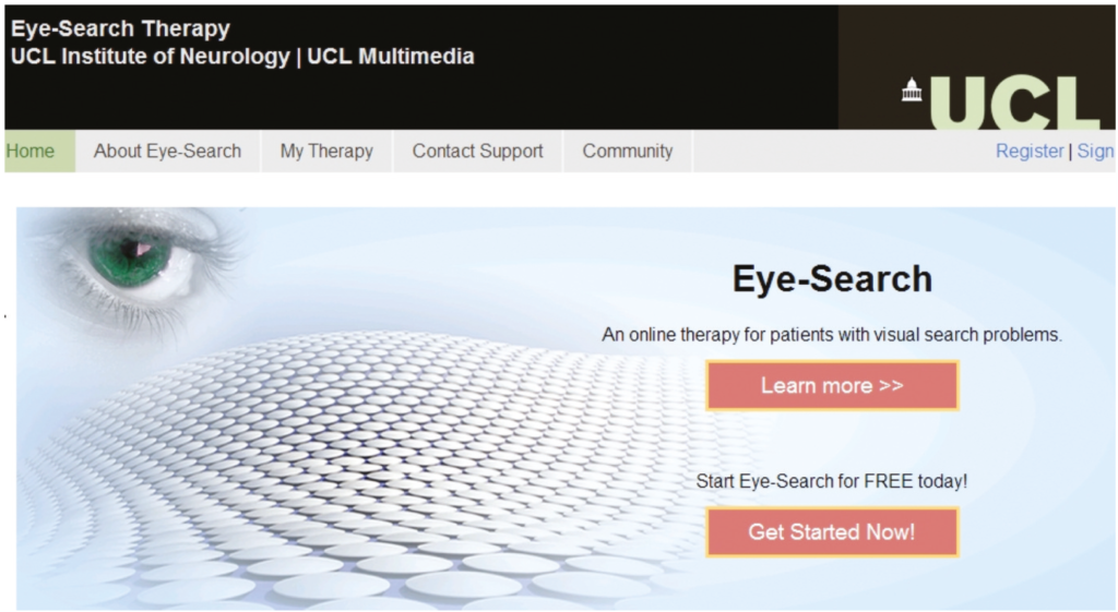
There are two, free-to-use, web-based therapies for patients with hemianopia for patients with either: 1) hemianopic alexia: http://www.readright.ucl.ac.uk; or, 2) problems with visual search: http://www.eyesearch.ucl.ac.uk (Figure 1). Both contain testing and therapy materials and are designed for home use by patients. Evidence for the efficacy of the Read-Right diagnostic and therapy tools has been published [14,15].
Prism adaptation for hemianopia, which can be successfully used to improve navigation within the environment [16], is anecdotally poorly tolerated by patients as any gains in one part of the visual field are at the expense of losses to another; also, the prisms required are very different to those used to correct double vision and there are very few NHS providers.

‘Higher’ visual function (pure alexia, neglect)
The evidence for therapeutic interventions in more complex perceptual disorders (pure alexia, prosopagnosia, simultanagnosia) is sporadic and weak and it is difficult to make any recommendations for specific therapies, although cross-modal therapy for pure alexia looks promising [17]. Spatial neglect or inattention is more common and has a stronger evidence base although the presumed efficacy for the main therapy (prism adaptation) has been questioned in a well conducted review [18]. Drug therapies have also been tried, with a recent study showing a small (10%) but significant improvement in table-top tests of selective attention when transdermal rotigotine, a dopamine agonist, was compared against placebo [19].
Summary
The therapeutic landscape for patients with language and/or visual impairments post-stroke is improving rapidly. There are many completed and ongoing small-scale trials of adjuncts that aim to improve the efficiency of rehabilitation: pharmacological and brain stimulation techniques. Rehabilitation of adult cognition can be viewed as a form of learning or re-learning and as such, whatever the role of exciting new adjuncts, requires a certain amount of brute-force in terms of practice-based learning. The most pertinent issue now is not whether these therapies work but how best to deliver them in a high enough dose so that patients can make meaningful gains? Self-supporting internet-based therapies seem the ideal solution for many specific therapy programmes.
References
- Price, CJ. The anatomy of language: a review of 100 fMRI studies published in 2009. Ann N Y Acad Sci 2010;1191:62-88. https://doi.org/10.1111/j.1749-6632.2010.05444.x
- Bowen, A, Hesketh, A, Patchick, E, Young, A, Davies, L, Vail, A, et al. Effectiveness of enhanced communication therapy in the first four months after stroke for aphasia and dysarthria: a randomised controlled trial. BMJ 2012;345:e4407. https://doi.org/10.1136/bmj.e4407
- Brady, MC, Kelly, H, Godwin, J, & Enderby, P. Speech and language therapy for aphasia following stroke. Cochrane Database of Systematic Reviews 2012(5). https://doi.org/10.1002/14651858.CD000425.pub3
- Bhogal, SK, Teasell, R, & Speechley, M. Intensity of aphasia therapy, impact on recovery. Stroke 2003;34(4):987-93. https://doi.org/10.1161/01.STR.0000062343.64383.D0
- Code, & Heron, C. Services for aphasia, other acquired adult neurogenic communication and swallowing disorders in the United Kingdom, 2000. Disabil Rehabil 2003;25(21):1231-7. https://doi.org/10.1080/09638280310001599961
- Seniow, J, Litwin, M, Litwin, T, Lesniak, M, & Czlonkowska, A. New approach to the rehabilitation of post-stroke focal cognitive syndrome: effect of levodopa combined with speech and language therapy on functional recovery from aphasia. J Neurol Sci 2009;283(1-2):214-8. https://doi.org/10.1016/j.jns.2009.02.336
- Crinion, J. Shocking speech. Aphasiology 2012;26(9):1077-81. https://doi.org/10.1080/02687038.2012.714215
- Lee, S, Shafe, AC, & Cowie, MR. UK stroke incidence, mortality and cardiovascular risk management 1999-2008: time-trend analysis from the General Practice Research Database. BMJ Open 2011;1(2):e000269. https://doi.org/10.1136/bmjopen-2011-000269
- Moss, A, & Nicholas, M. Language rehabilitation in chronic aphasia and time postonset: a review of single-subject data. Stroke 2006;37(12):3043-51. https://doi.org/10.1161/01.STR.0000249427.74970.15
- Zhang, X, Kedar, S, Lynn, MJ, Newman, NJ, & Biousse, V. Natural history of homonymous hemianopia. Neurology 2006;66(6):901-5. https://doi.org/10.1212/01.wnl.0000203338.54323.22
- Warren, M. Pilot study on activities of daily living limitations in adults with hemianopsia. Am J Occup Ther 2009;63(5):626-33. https://doi.org/10.5014/ajot.63.5.626
- Pollock, A, Hazelton, C, Henderson, CA, Angilley, J, Dhillon, B, Langhorne, P, et al. Interventions for visual field defects in patients with stroke. Cochrane Database of Systematic Reviews 2011(10). https://doi.org/10.1002/14651858.CD008388.pub2
- Schuett, S, Heywood, CA, Kentridge, RW, Dauner, R, & Zihl, J. Rehabilitation of reading and visual exploration in visual field disorders: transfer or specificity? Brain 2012;135(Pt 3):912-21. https://doi.org/10.1093/brain/awr356
- Koiava, N, Ong, YH, Brown, MM, Acheson, J, Plant, GT, & Leff, AP. A ‘web app’ for diagnosing hemianopia. J Neurol Neurosurg Psychiatry 2012. https://doi.org/10.1136/jnnp-2012-302270
- Ong, YH, Brown, MM, Robinson, P, Plant, GT, Husain, M, & Leff, AP. Read-Right: a “web app” that improves reading speeds in patients with hemianopia. J Neurol 2012. https://doi.org/10.1007/s00415-012-6549-8
- Bowers, AR, Keeney, K, & Peli, E. Community-based trial of a peripheral prism visual field expansion device for hemianopia. Arch Ophthalmol 2008;126(5):657-64. https://doi.org/10.1001/archopht.126.5.657
- Woodhead, ZVJ, Penny, W, Barnes, GR, Crewes, H, Wise, RJS, Price, CJ, et al. Reading therapy strengthens top-down connectivity in patients with pure alexia. Brain 2013 (in press). https://doi.org/10.1093/brain/awt186
- Barrett, AM, Goedert, KM, & Basso, JC. Prism adaptation for spatial neglect after stroke: translational practice gaps. Nature Reviews Neurology 2012;8(10):567-77. nhttps://doi.org/10.1038/nrneurol.2012.170
- Gorgoraptis, N, Mah, YH, Machner, B, Singh-Curry, V, Malhotra, P, Hadji-Michael, M, et al. The effects of the dopamine agonist rotigotine on hemispatial neglect following stroke. Brain 2012;135:2478-91. https://doi.org/10.1093/brain/aws154
