Abstract

François Pourfour du Petit (1664-1741) was a Parisian experimental Neuroanatomist, and Ophthalmologist.
Based on his extensive experiences of brain and spinal injuries as a military doctor in the armies of Louis XIV he performed many animal experiments that demonstrated the anatomy and functional significance of the cervical sympathetic nerves, correcting previous errors of Thomas Willis and Raymond Vieussens.
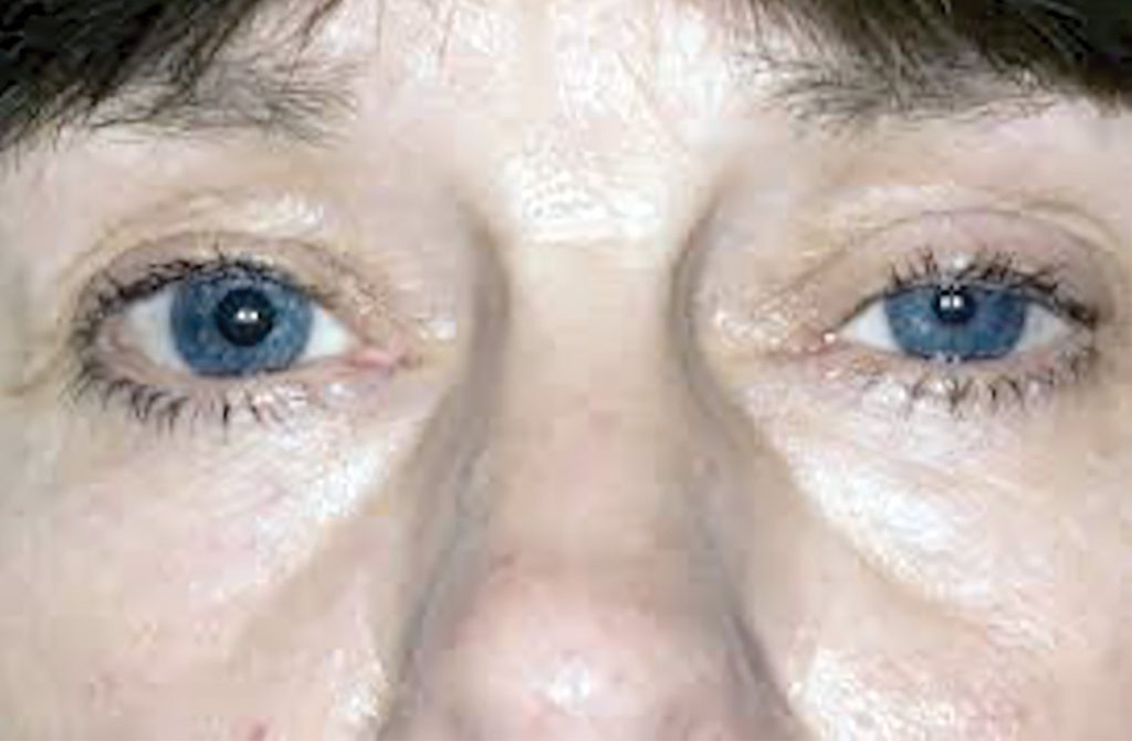
He long predated the descriptions of Horner’s syndrome (1869) when he showed that interruption of sympathetic pathways inactivated both the dilator muscle and produces miosis, and the superior tarsal muscle, which produces ptosis and enophthalmos. This was later elaborated by Hare, Weir Mitchell and Claude Bernard.
The tetrad of ptosis, miosis, enophthalmos, and impaired facial sweating was described in 1869 by Johann Friedrich Horner (1831-1886) (Figure 1), Professor of Ophthalmology in Zurich:
Anna Brändli, aged 40, a healthy looking peasant woman…six weeks after her last confinement noticed a slight drooping of her right upper eyelid, …The pupil of the right eye is considerably more constricted than that of the left, but reacts to light; the globe has sunk inward very slightly…During the clinical discussion…the right side of her face became red and warm; while the left side remained pale and cool. …The patient thereupon told us that the right side had never perspired,… the thermometer on the right recorded 35°C. that on the left, 30°C.…” [1,2]
Horner’s syndrome (oculosympathetic paresis) had been described several times before his report. Edward Selleck Hare, in 1838, and Silas Weir Mitchell both gave earlier accounts: Hare’s in a cervical tumour, Weir Mitchell’s in a gunshot neck wound [3].

The sympathetic nerves
Long before these descriptions, the anatomy and function of the sympathetic chain had been studied but mistakenly represented. It is easily forgotten that at this time there was little scientific or rational scientific medicine. The era was of magic, miasma and witchcraft. Insubstantial ideas of the spiritus animalis were rife, and irrational speculation abounded.
The sympathetic chain was known as the intercostal nerve, a name introduced by Thomas Willis (1621–1675). He described:
The principal trunk of the intercostal nerve as consisting of two or three twigs, which were sent back from the fifth and sixth nerves, and joined together as in one stem [4].
Thus he believed the sympathetic descended from these cranial nerves along the rib cage: hence his term intercostal. Similarly, Raymond Vieussens (c.1641-1715), a distinguished anatomist and pathologist from Montpellier and physician to the Hospital of St. Eloys described the ophthalmic branch of the fifth cranial nerve as:
Emitting, sometimes one, sometimes two fibres, which with a fibre emitted from the sixth cranial nerve became attached to the intercostal nerve [5,6].

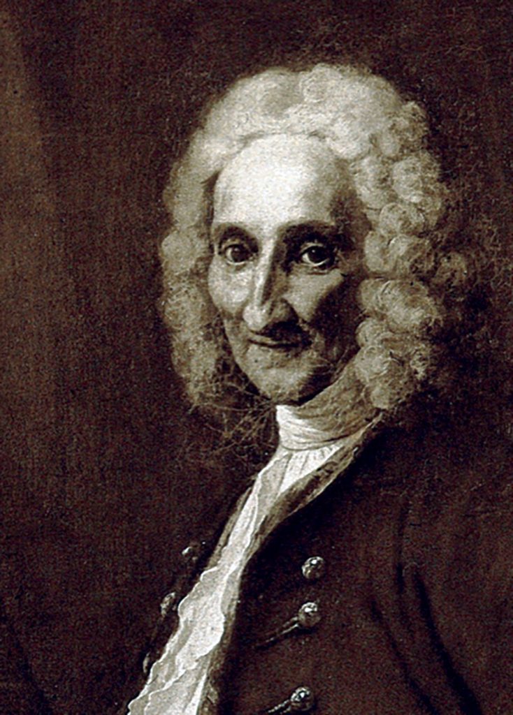
François Pourfour du Petit (1664-1741) (Figure 2), a Parisian experimental Neuroanatomist, and Ophthalmologist refuted these accepted ideas. He had scrutinised and investigated his extensive experiences of brain and spinal injuries as military doctor in the armies of Louis XIV between 1693 and 1713. He verified his conclusions by many animal experiments. In this way he demonstrated the anatomy and functional significance of the cervical sympathetic chain.
In 1727 Du Petit described the intercostal nerve as ascending, entering the skull along with the carotid artery in the twisted petrous tube [7]. Crucially, he recognised the preganglionic fibres arising from T1 to L3 spinal segments. Thus, he disproved both Willis and Vieussen’s identification of the origin of the sympathetic chain in the fifth and sixth cranial nerves. Although du Petit’s results were unequivocal, lamentably they remained latent, largely ignored until the nineteenth century.
In 1727, he reported that cutting the superior sympathetic nerves of dogs produced ptosis and miosis [7].
His later dog experiments convinced him that miosis was due to connections between the ciliary nerves with the intercostal nerves. He showed that interruption of sympathetic pathways (oculosympathetic paresis) inactivates both the dilator muscle and produces miosis, and the superior tarsal muscle, which produces ptosis and the appearance of enophthalmos. This contrasted with third nerve palsy where ptosis and a dilated pupil result from a loss of innervation to the sphincter pupillae.
In a soldier whose neck had been slashed by a sword he described dilatation of the pupil (mydriasis) eyelid retraction, and hemifacial hyperhidrosis – the syndrome of Pourfour Du Petit – in effect a reverse Horner’s syndrome. He experimentally confirmed these signs in dogs by cutting their cervical sympathetic chain.
He also showed with originality that brain injuries could cause weakness or paralysis of the contralateral limbs, and he established the medullary decussation of the pyramidal tracts.
Pourfour du Petit-Claude Bernard-Horner syndrome
There are few eponyms more clumsy and confusing than the ‘Pourfour du Petit-Claude Bernard-Horner syndrome’, each with disputed demands for priority [8]. The history of Petit’s clinical observations and experiments, though thoroughly recorded by Best [6], failed to achieve recognition. In 1851 Claude Bernard using rabbits repeated and confirmed his conclusions and gave his own distinctive description [9].
The nomenclature unsurprisingly proved polemical. Paul Bonnet’s paper [10] fiercely defended the eponym of Pourfour du Petit instead of Horner:
Il est temps que le nom de Horner – tout au moins en ce qui concerne le syndrome paralytique du sympathique – rentre dans l’ombre, d’ou il n’aurait jamais du sortir”. [It is time that the name of Horner – at least with respect to the paralytic syndrome of the sympathetic – returns to the shadows, which it should never leave again].
Of the original run of 200 copies of du Petit’s pamphlet – Lettres d’un medecin des hôpitaux du roy, a un autre medecin de ses amis – only three copies remain. This may explain why citations to his work are sparse. One copy is in the Bibliotheque Nationale, and there was a “London copy” photographed by William Osler [11].
Sympathetic denervation hypersensitivity in the first, second, or third order (pre-postganglionic) fibres can be shown with the aid of cocaine and adrenaline installations, to localise the lesion. The many causes are disclosed by associated neurological symptoms and neuroradiology.
Whereas du Petit and Claude Bernard experimented mainly in animals, reference to oculosympathetic paralysis in man was recorded but uncommon before Horner. For his detailed clinical account he deserves full credit.
References
- Horner JF; Über eine Form von Ptosis. Klinische Monatsblätter für Augenheilkunde, Stuttgart, 1869, 7: 193-198. Translated by Fulton JF. Horner And The Syndrome Of Paralysis Of The Cervical Sympathetic. The Archives Of Surgery April, 1929; 18:2025-2039. https://doi.org/10.1001/archsurg.1929.01140131129078
- Fulton JF. Horner and the syndrome of paralysis of the cervical sympathetic. Arch Surg 1929; 18: 2025-39. https://doi.org/10.1001/archsurg.1929.01140131129078
- Pearce JMS. A note on Claude Bernard-Horner’s syndrome. J Neurol Neurosurg Psychiatry. 1995; 59(2): 188, 191. https://doi.org/10.1136/jnnp.59.2.188
- Wilis T. Cerebri anatome, cui acessit Nervorum descriptio et usus, Typis Tho. Roycroft, Impensis Jo. Martyn. London,1664; p.184. [plates by Christopher Wren and Richard Lower.]
- Vieussens R. Neurographia Universalis, Lyons, 1684, p. 170.
- Best AE. Pourfour du Petit’s experiments on the origin of the sympathetic nerve. Med Hist. 1969;13(2):154-74. https://doi.org/10.1017/S0025727300014253
- Pourfour du Petit F. Memoire dans lequel il est d que les nerfs intercostaux fournissent des rameaux que portent les esprits dans les yeux. Hist Acad Roy Sci (Paris) 1727: 1-19. Cited by Best. (ref 5.)
- Bruyn GW, Gooddy W. Horner’s syndrome. In: Neurological Eponyms edited by Peter J. Koehler, George W. Bruyn, John M. S. Pearce. New York, Oxford, OUP. 2000. pp.227-233.
- Bernard, Claude. Influence du grand sympathique sur la sensibilité et sur la calorification. C. R. Soc. Biol., t. 3, 1851 (1852), pp. 163-164
- Bonnet P. L’histoire du syndrome de Claude Bernard. Le syndrome paralytique du sympathique cervical. Arch d’Ophtalm (Paris) 1957;17:121-38.
- Kruger L., Swanson L.W. (2007) 1710: The Introduction of Experimental Nervous System Physiology and Anatomy by François Pourfour du Petit. In: Whitaker H., Smith C.U.M., Finger S. (eds) Brain, Mind and Medicine: Essays in Eighteenth-Century Neuroscience. Springer, Boston, MA. https://doi.org/10.1007/978-0-387-70967-3_8