Abstract
This paper recalls the descriptions and early ideas about the dilated pupil accompanying raised intracranial pressure resulting from head injuries and space-occupying lesions. The observation of the ominous fixed, dilated pupils in those with expanding brain lesions dates to Richard Bright and Jonathan Hutchinson in the 19th century, but its significance and mechanisms were only debated in the early years of the 20th century. Compression or stretching of the oculomotor nerve were considered possible causes, but the related mechanisms of coning and the importance of lateral shift were only more recently realised.
FEW investigations of late years have excited more interest than those which have been made into the connection of certain changes in the eye with diseases of the central nervous system, and into the additional means of diagnosis which such changes may afford. Sir Thomas Clifford Allbutt ,1872
Not until the early 20th century was the importance of a fixed dilated pupil recognised as an ominous physical sign. It became a neurosurgical axiom that a fixed dilated pupil occurs ipsilateral to a pressure cone caused by a space-occupying lesion with intracranial hypertension [2].
Brain herniations are traditionally classed as: subfalcial, uncal (transtentorial), and cerebellar tonsillar. They can complicate head injury or any other causes of a brain mass or swelling [3]. Central herniation, usually preceded by uncal and cingulate herniation, is the downward movement of the brain through the tentorial notch. Clinically it is manifested by stupor leading to coma; small, reactive pupils becoming fixed and dilated; with irregular breathing, leading to decerebrate posture, and ultimately death. With herniation, the ipsilateral posterior cerebral artery may be compressed adding to the ischaemia induced by brain oedema.
Observations of raised intracranial pressure, and more recently papilloedema (in early accounts described as ‘choked disc’, stauung-spapille, or optic neuritis) were recorded more than a century ago [1,4]. But neither the mechanisms nor clinical significance of the associated dilated pupil were fully understood.

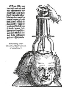
The redoubtable surgeon, Percivall Pott (1714-1788) detailed his Observations on the Nature and Consequences of those Injuries to which the Head is liable from external Violence [5] and referred to the woodcuts of the surgeon Hans von Gersdorff (c.1455-c.1529). But Pott failed to mention the ocular signs in von Gersdorff’s ancient woodcut, the work of Johannes [syn. Hans] Wechtlin, contained in his Feldbuch der Wundartzney (Fieldbook of wound medicine) 1517. This illustrated the elevation of a depressed skull fracture. The patient shows a slightly dilated right pupil and the eye is abducted, suggesting a partial third nerve palsy (Figure 1). It has been said that his observation was not repeated for another 300 years. However, in this oft-cited instance [6] there is insufficient evidence to wholly exclude unrelated strabismus with a physiological asymmetry of the pupils of up to 0.4 mm, found in about 20% of normal people.
John Cheyne (1777-1836), remembered for Cheyne-Stokes respiration, studied medicine at Edinburgh, where Alexander Monro secundus (1733-1817), who described the interventricular foramen, was one of his teachers. In cases of apoplexy, Cheyne believed that cerebral anaemia might be the cause; in an early case in 1812 he recorded the contraction and dilatation of the pupil and noted, ‘Thus we do not despair until the pupil ceases to contract.’ [7]
Richard Bright

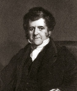
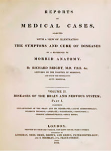
A more comprehensive description is found in 1831, in Volume two* of Richard Bright’s (1789-1858) (Figure 2) famous case reports [8] devoted to neuropathology [9] with 54 magnificent plates (Figure 3). William Munk in his Roll was justifiably dazzled by Bright’s abilities:
‘Dr. Bright showed the most sagacious observation, untiring industry, and wonderful powers of investigating truth, the end and aim of all his work [10].’ Bright reported the ipsilateral dilated pupil in a man whose post-mortem showed an epidural haematoma with petechial haemorrhages in the brain after head injury (case 191) [8,11]. On the second day after a well-described lucid interval, the patient was noted to be less responsive and to have a slow pulse. Bright observed:
A 38-year-old man working at a large wharf below London bridge fell from a height of 11 or 12 feet. The following day his language was incoherent and speech scarcely articulate, and he complained of pain in the head. He was bled, and on the third day he is in a state of stupor, but may be roused to answer question. The muscles of the left side of the face are paralyzed… right pupil dilated. He died the sixth day and an autopsy was performed. An epidural hematoma was found. In general the pupils sometimes contracted, at other times dilated, and acting quite irregularly under the stimulus of light.
Bright described another case, a 20-year-old man with apoplexy whose pupils dilated, and did not contract when a candle was brought near. He clearly recognised the importance of this vital clinical sign; however, he suggested no mechanism.

Bright’s salient observation may have prompted the experiments of Ernst Viktor von Leyden (1832-1910), which in 1866 showed that with increased intracranial pressure, the pupils first became narrow, then dilated, but not always symmetrical, accompanied by coma with a slow pulse, impaired respiration and death [12].Five years later, Alexander Pagenstecher (1828-79) studied consecutive reactions of the pupil with increasing experimental pressure, noting an initial constriction followed by a dilated fixed pupil with a stupor or coma [13,14]. The controversial Guy’s hospital surgeon and anatomist Astley Paston Cooper (1768-1841) also experimented on dogs, manually compressing the exposed dura, trying to distinguish compression from ‘simple concussion’. In humans he explained the symptoms caused by compression [15]: (lecture XVII):
The breathing being stertorous, the pulse slow, and the pupils dilated; …when you then find a patient with the apoplectic stertor, slow pulse, dilated pupils, it will generally happen that the brain is compressed.
Hutchinson’s pupil
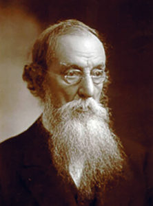
It remained for Jonathan Hutchinson (1828-1913)16 (Figure 4) in 1867 to provide a more detailed report of his experience with human head injuries and his observations of a dilated pupil on the side of a fatal traumatic epidural haematoma [17]:
…Unilateral dilatation of pupil after injury to the head. A man had died in whom this symptom was present, and in whom we found a large clot of blood between the dura mater and the bone pressing forwards upon the sphenoidal fissure, and no doubt compressing the trunk of the third nerve. …We can have little hesitation in assuming that the cause of death was compression of the brain.
Hutchinson also reported a boy, who was admitted on a Thursday, having been knocked down in the street and possibly run over. He did not relate the dilated pupil to transtentorial shift, but invoked the mechanism of third nerve compression that affects the ocular para-sympathetic causing the pupil to dilate, and to fail to constrict in response to light:
From the position of the clot there can be little doubt that the third nerve is compressed and thus, the dilatation of the pupil is explained. These two cases, so exactly parallel, seem to supply us with a new and very valuable symptom indicative of effusion of blood in this situation. … You will see that beyond the symptoms which usually attend cases of severe concussion of the brain (accompanied as they frequently are by more or less of contusion also), we had had none excepting the dilatation of the right pupil. The boy had been conscious up to within about half an hour of his death; he had had a rapid pulse and great restlessness throughout; he had had no observable paralysis [17].

The Guy’s hospital surgeon, WHA Jacobson (1847-1924) suggested in 1886, that this phenomenon should be named ‘Hutchinson pupil’ [18]. He may have neglected Bright’s earlier account.
William Macewen (1848–1924) in 1887 considered pupillary dilatation to be a sign of oculomotor nerve irritation, paralysis or vascular changes in connection with a middle cranial fossa clot [19].
Duret Haemorrhage
Henri Duret (1849–1921) a surgeon, who trained with Charcot and Vulpian [20], reported that blows on the head in animals increased intracranial pressure. He observed loss of consciousness, rigidity, slow then faster breathing, and mid-dilated pupils; days later the pupils constricted and the animal died. In addition to the cerebral damage at autopsy he observed ‘haemorrhagic lesions in the superior part of the bulbar base’, which proved ‘that the blows to the skull may have a considerable effect on the bulbus’ [21,22]:
A dotted line of haemorrhages on the floor of the medulla’s thickness and around the central canal… this is explained by the fact that at the time of impact, the fluid in the ventricles has an effect on the cerebral aqueduct, the fourth ventricle and, especially, the spine’s central canal.
Mistakenly, he considered:
the sudden cessation or suppression of brain function subsequent to an impact to the skull is produced by means of the cerebrospinal fluid, which transmits the damaging action to regions of the brain capable of generating all the observed phenomena.
This mechanism he termed choc céphalo-rachidien (cephalospinal shock). He distinguished two stages, first, pupil constriction by bulbar lesions due to choc céphalo-rachidien, secondly, dilatation from the accumulation of blood around the oculomotor nerve. Theodor Kocher (1841–1917), a Swiss surgeon then used the eponym ‘Duret haemorrhages’ in his comprehensive review of brain injuries [23].
Later investigations
The German surgeon, Ernst von Bergmann (1836–1907), a keen advocate of Lister’s aseptic techniques, described fixed dilated pupils in his text on brain injuries [24]. He observed that the ipsilateral pupil became narrower at first and then, with increased pres-sure leading to coma, it dilated.
In 1904 James Collier observed both the cerebellar pressure cone and ‘false localising signs’ in cases of intracranial tumour examined clinically and pathologically [25]. He distinguished this from the dilated pupil of uncal herniation [26]. He commented [27]:
In many cases of intracranial tumour of long duration, it was found post-mortem that the posterior inferior part of the cerebellum had been pushed down and backwards into the foramen magnum and the medulla itself somewhat caudally displaced, the 2 structures together forming a cone-shaped plug tightly filling up the foramen magnum.
Adolf Meyer (1866-1950) related the pressure on the third nerve to herniation of the uncus into the incisura angularis [28]. Similarly, Sir Geoffrey Jefferson (1886-1961) described in four cases the mechanism of temporal lobe herniation: a pressure cone formed by uncal herniation on the crus cerebri, with resulting impingement on the oculomotor nerve [3].
Later accounts of Kernohan-Woltman’s notch of the crus cerebri causing ipsilateral hemiplegia [29] were followed by the newer concept that depression of alertness corresponded to distortion of the brain by horizontal rather than vertical displacement.
Holman and Scott pointed out that although a dilated pupil was valuable in locating the site of injury, there were only a few descriptions in the literature of patients who deteriorated and lapsed into a coma.
The mechanism of its appearance is not obvious, but it is assumed that the intracranial course of the third nerve, as it lies against the bony wall of the cranium, lends itself peculiarly well to compression from a pressure applied lateral and superior to it [30].
This mirrored Hutchinson’s cautionary comment:
This case adds another to a considerable series which we have recently had showing that the orthodox symptoms of compression of the brain, absolute insensibility, stertor, slow, laboured pulse, and hot surface, are not by any means always met with… [17]
Holman and Scott echoed Horsley’s earlier report [4] of the consequences of this sign for surgery:
‘[We] place reliance on the unilateral dilatation and fixation of the pupil as an indication of unilateral cerebral compression, due more particularly to haemorrhage…Unilateral dilatation and fixation of the pupil is a valuable aid in determining the location of the intracranial injury and haemorrhage following head injuries. When one pupil was dilated the operation should be on the ipsilateral side.‘ [30]
Miller Fisher
With characteristic shrewdness, in 1995 Miller Fisher (1913-2012) investigated transtentorial herniation with computed tomography: descent through the tentorial opening could not be documented. Stressing the importance of lateral displacement, he warned that bilateral brain stem compression in acute bilateral cases must be distinguished from herniation:
Upward cerebellar herniation is only the sign of an overfull posterior fossa. …Subfalcial herniation is tolerated unless lateral displacement is excessive…Combining clinical, pathologic, computed tomography and magnetic resonance imaging data, it is concluded that temporal lobe herniation is not the means by which the midbrain sustains irreversible damage in acute cases, but rather lateral displacement of the brain at the tentorium is the prime mover and herniation a harmless accompaniment [31]. The dilated pupil is attributed to midbrain distortion instead of uncal herniation.
Conclusion
It is scarcely necessary to conclude that in this context the mechanisms of abnormal pupils remain debatable since many fundamental questions still await solution. The unilaterally dilated pupil can be explained by stretching of the oculomotor nerve over the clivus or by its compression by a bulging hemispheric mass or haemorrhage. It appears that depression of consciousness is mainly related to a lateral brain shift at the tentorium. The false-localising impaired pupil reaction on the opposite side is attributed to midbrain damage due to distortion and compression [32,25].
References
- Allbutt TC. On the causation and significance of the choked disc in intracranial diseases. Br. Med. J. 1872;1:443-445. https://doi.org/10.1136/bmj.1.591.443
- Marshman LAG, Polkey CE, C. Penney CC. Unilateral Fixed Dilation of the Pupil as a False-localizing Sign with Intracranial Hemorrhage: Case Report and Literature Review. Neurosurgery 2001;49:1251-6. https://doi.org/10.1227/00006123-200111000-00045
- Jefferson G: The tentorial pressure cone. Arch Neurol Psychiatry 1938;40:857-876. https://doi.org/10.1001/archneurpsyc.1938.02270110011001
- Horsley V. “Optic Neuritis,” or ” Papilloedema.” Treatment, Localizing Value, and Pathology. Br. Med. J. 1910;1:553-558. https://doi.org/10.1136/bmj.1.2566.553
- Pott P. Observations on the Nature and Consequences of those Injuries to which the Head is liable from external Violence, London. L. Hawes, W. Clarke, & R. Collins 1768.
- Flamm ES. The dilated pupil and head trauma 1517-1867. Med Hist 1972;16:194-199. https://doi.org/10.1017/S0025727300017610
- Cheyne J. Cases of Apoplexy and Lethargy: With Observations Upon the Comatose Diseases. London: Underwood, 1812. https://archive.org/stream/casesofapoplexyl00chey/ casesofapoplexyl00chey_djvu.txt
- Bright R. Reports on Medical Cases selected with a View of illustrating the Symptoms and Cure of Diseases by a Reference to Morbid Anat. Vol 2. London, Longman, 1831.
- Pearce JMS. Richard Bright and His Neurological Studies. Eur Neurol 2009;61:250-254. https://doi.org/10.1159/000198419
- Munk’s Roll. Lives of the Fellows RCP London. Richard Bright. Volume III, page 155.
- Pearce JMS. Observations on Concussion. Eur Neurol 2008;59:113-119 https://doi.org/10.1159/000111872
- Von Leyden E. Beitrage und Untersuchungen zur physiologie und pathologie des gehirns. Virchow’s archives 1866;37:519- 59. Cited by Eelco FM Wijdicks In: Famous First Papers for the Neurointensivist. New York: Springer, 2013.12-16 https://doi.org/10.1007/BF01935598
- Pagenstecher F: Experimente und Studien über Gehirndruck. Heidelberg: Winter, 1871 Cited by Koehler PJ and Wijdicks EFM. Ref 14
- Koehler PJ, Wijdicks EFM. In: Fixed and dilated: the history of a classic pupil abnormality. J Neurosurg 2015;122:453-463. https://doi.org/10.3171/2014.10.JNS14148
- Cooper A. The Principles and Practice of Surgery. Lee A, ed. London: E Cox, 1836. Pp. 160-161.
- Pearce JMS. Sir Jonathan Hutchinson and sarcoidosis: A brief history. Ann R Coll Surg Engl 2010;92(8)280-82. https://doi.org/10.1308/147363510X12779829074860
- Hutchinson J. Four lectures on compression of the brain, Clinical Lectures and Reports of the London Hospital. 1867- 1868,4,10-55.
- Jacobson,W.H.A. On middle meningeal haemorrhage. Guy’s Hospital Reports 1885;(6)43:147-308
- Macewen W. The pupil in its semiological aspects. Am J Med Sci 1887;94:123-146. https://doi.org/10.1097/00000441-188707000-00007
- Walusinski O, Courrivaud P. Henry Duret (1849-1921): A Surgeon and Forgotten Neurologist. Eur Neurol 2014;72:193- 202. https://doi.org/10.1159/000361046
- Duret H. Etudes expérimentales sur les trau- matismes cérébraux. Thèse, Paris, 1878, n° 64, Versailles, Imprimerie Cerf, 1878, p 339.
- Duret H. On the role of the dura mater in cerebral traumatism. Brain 1878;1:29-47. https://doi.org/10.1093/brain/1.1.29
- Kocher T. Hirnerschütterung, Hirndruck und chirurgische Eingriffe bei Hirnkrankheiten. Specielle Pathologie und Therapie. Ed, Nothnagel H. Vienna: Hölder, 1901; Vol 9.
- Von Bergmann E.Die Chirurgische Behandlung der Hirnkrankheiten (1888; “The Surgical Treatment of Brain Disorders”).
- Larner AJ. False localising signs Journal of Neurology. Neurosurgery & Psychiatry 2003;74:415-418. https://doi.org/10.1136/jnnp.74.4.415
- Pearce JMS. James Collier (1870-1935) and uncal herniation. J Neurol Neurosurg Psychiatry. 2006; 77(7): 883-884. https://doi.org/10.1136/jnnp.2006.087544
- Collier J. The false localizing signs of intracranial tumour. Brain 190427490-508.
- Meyer A. Herniation of the brain. Arch Neurol Psychiatry 1920;4:387-400. https://doi.org/10.1001/archneurpsyc.1920.02180220036003
- Kernohan J, Woltman H. “Incisura of the crus due to contralateral brain tumour”. Arch Neurol Psychiatry. 1929;21: 274-287. https://doi.org/10.1001/archneurpsyc.1929.02210200030004
- Holman E, Scott WMJ. Significance of unilateral dilatation and fixation of pupil in severe skull injuries. JAMA. 1925;84:1329-32. https://doi.org/10.1001/jama.1925.02660440015006
- Fisher CM. Brain Herniation: A Revision of Classical Concepts. Can. J. Neurol. Sci. 1995; 22: 83-91. https://doi.org/10.1017/S0317167100040142
- Ropper AH. The opposite pupil in herniation. Neurology 1990;40:1707-9. https://doi.org/10.1212/WNL.40.11.1707