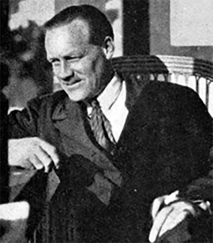
The harmless condition of a dilated pupil which reacts abnormally slowly (myotonic) to light and convergence was fully described in 1931 by William John Adie (1886-1935) [1], of the National Hospital, Queen Square:
I wish to draw attention to a benign symptomless disorder characterized by pupils which react on accommodation but not to light, and by absent tendon reflexes. Five of the six cases I am about to describe came under my notice in the course of a few weeks;… Though harmless in itself it merits recognition because it is often mistaken for a manifestation of syphilis of the nervous system, with unfortunate consequences… Mr. Foster Moore has described seven cases under the title ‘Non-luetic Argyll Robertson pupil. [2]
Usually seen in females, in 80% of cases it is unilateral. Adie was not the first to observe this uncommon curiosity and its variant manifestations. In 1818 the London ophthalmologist, James Ware (1756–1815), described a patient who may have had a myotonic pupil [3]. His patient, whose pupillary abnormality had been known for twenty years, was:
A lady between thirty and forty years of age, the pupil of whose right eye, when she is not engaged in reading, or in working with her needle, is always dilated very nearly to the rim of the cornea; but whenever she looks at a small object, nine inches from the eye, it contracts, within less than a minute, to a size nearly as small as the head of a pin. Her left pupil is not affected like the right; but in every degree of light and distance, it is contracted rather more than is usual in other persons.… Several instances have come under my notice, in which the pupil of one eye has become dilated to a great degree, and has been incapable of contracting on an increase of light.
Adie stated: ‘The tonic or myotonic pupillary reaction was first described in 1902 by Saenger in the left pupil of a 34-year-old woman) [4] and by Strasburger in a 17-year-old man [5] independently [6]. Later in the same year both Saenger and Nonne reported further cases for which Saenger proposed the name ‘myotonic pupillary movement’ to distinguish it from other forms of mydriasis or abnormal pupillary reflexes. Later, Adie related ‘atypical ‘forms, less clearly defined, but he included some forms of ‘internal ophthalmoplegia and complete light rigidity with atonic convergence reaction.’
Before Adie, Hughlings Jackson in 1881 had also clearly described the same disorder [7]:
A woman aged 26 was sent to see me simply because the right pupil was much larger than the left. It had been so three years…the right pupil was dilated and absolutely motionless to light, and also during accommodation, yet the accommodation itself on this side was perfect; this was severely tested by Mr Couper…this case at first puzzled me…It occurred to me to test the knees. Neither I nor Mr Couper found the smallest trace of the knee phenomenon [knee jerk]. Several times did I pertinaciously inquire for other symptoms of tabes; there were no other symptoms of any kind.…Dr Buzzard …confirmed the above observations. [8]
In 1906 Markus had described an isolated case of partial iridoplegia [9], and Weill and Reys [10] also described tonic pupil reactions to convergence and accommodation with areflexia. Some reports bear their names as eponyms.
Adie’s more detailed, classic paper in 1932 [11], described 22 patients, with absent tendon reflexes, and noted 44 reported cases of tonic pupil in nine of which there were absent tendon reflexes. In this account he outlined four incomplete forms (the last would not now be accepted):
1. The complete form—typical tonic pupil and absence of reflexes.
2. Incomplete forms : a) tonic pupil alone; b) atypical phase of the tonic pupil alone (iridoplegia; internal opthalmoplegia); c) atypical phases of the tonic pupil with absent reflexes; d) absent reflexes alone.
At about the same time, Gordon Holmes found 19 patients with the myotonic pupils, characterised in his Introduction to Clinical Neurology:
By very slow contraction on convergence, and even slower relaxation. The reflex to light is often lost too. One or both eyes may be affected.
Holmes’s 1931 paper described it and its association with symptoms of other diseases of the nervous system:
Frequently no change in the size of the pupil was visible immediately on convergence, but when this was maintained for a few seconds the pupil slowly and gradually grew smaller, till its diameter equalled or was even narrower than that of the normal eye. The rate of contraction varied very much…When contracted the pupil remains constant and when convergence is relaxed it dilates slowly… In the present state of our knowledge a separation of those cases in which the tendon jerks are absent from those in which they persist is unjustifiable . . . the similarity of the symptoms in all these cases naturally suggests a common aetiology. [12]
Edwin Bramwell linked Holmes’s name with Adie’s in 1936 [13]. The brilliant George Bruyn mischievously pointed to the ‘peculiarity’ that Morgan, Symonds, Holmes, and Adie all published at about the same time, in different journals without referring to each other, yet they knew each other well at Queen Square [1]. However Adie’s second paper (1932) did mention Holmes’s work [11].
The credit must be Adie’s for stressing its harmless nature and crucially distinguishing it from neurosyphilis. Adie did not claim originality, recognising several earlier accounts. He acknowledged that Morgan and Symonds had recorded:
In Guy’s Hospital Reports for 1927 Dr Symonds and I drew attention to a small group of cases in which certain abnormalities of the pupil, including inequality and defective reaction to light and convergence, and also some affection of accommodation, were associated with a pathological absence or diminution of the tendon-jerks. [14]
It was previously confused with the Argyll Robertson pupil associated with luetic tabes dorsalis or General Paralysis of the Insane, characterised by: a loss of both direct and consensual light reflexes; pupillary inequality; irregularity and iris atrophy without reaction to light. Adie clearly distinguished this from his myotonic (Pseudo-Argyll Robertson) pupil.
Since Adie’s descriptions [2,6,11], this conception of an atypical tonic pupil has been widened [15], which unnecessarily complicates a diagnosis which is secure if clinical observations are precisely observed.
Pathogenesis

Stanley Graveson (1915-1976) studied 15 patients, three men, 12 women, aged 12 to 55 [16]. He set out to illuminate: (1) the site of the anatomical lesion and (2) the nature and specificity, or otherwise, of the underlying pathological process. Two types of tonic pupils were distinguished, (a) the fixed type, and (b) the ordinary type of tonic pupil. The only common features of this latter variety are (1) their regularity of shape or position of the pupil and (2) the slowness of pupillary dilatation after convergence. Graveson pointed out that the lesion had to be on the efferent limb of the light reflex arc, to account for the absence of light response in a unilateral tonic pupil with a simultaneous brisk consensual response in the normal pupil. The prompt reaction of the pupil to pilocarpine meant that the muscle of the iris could not be at fault.
The pathogenesis remained uncertain. Adie had reported: ‘all we can say is that the ocular and reflex signs we have considered seem to be the expression of a kind of perversion of nervous activity of which, at present, we can form no conception.’ Harriman and Garland, however, clarified matters in a minutely examined autopsy case in 1968 that showed the causal neuronal degeneration in the ciliary ganglion, denervation of the ciliary body, partial atrophy of pupilloconstrictor muscle – all on the right, affected side. The site of the lesion responsible for areflexia was less conclusive, but they noted a selective degeneration of neurones in dorsal root ganglia, possibly those supplying muscle spindles. This was consistent with a neurophysiological reduction in the size of the H waves, evidence of depression of the monosynaptic spinal reflex arc, previously observed [17]. It confirmed the similar complete loss of ciliary ganglion cells in the only other autopsied case at the time, [18] and fits with the known denervation cholinergic hypersensitivity of the pupil to methacholine and pilocarpine.
References
- Bruyn GW, Gooddy W. Adie’s syndrome. In: Neurological Eponyms. edited Peter J. Koehler PJ, Bruyn GW, Pearce JMS. Oxford, OUP 2000;181-185.
- Adie WJ. Pseudo-Argyll Robertson pupils with absent tendon reflexes. Br Med J 1931;1:1091. https://doi.org/10.1136/bmj.1.3676.1091
- Ware J. Observations relative to the near and distant sight of different persons. Phil Tr Roy Soc London 1813;103:36-38. https://doi.org/10.1080/14786441308638343
- Saenger A. Myotonic Pupil Movement .Neurol Centralbl 1902;21:837 and 1137.
- Strasburger J. Sluggishness of the Pupil to Accommodation and Convergence. Neurol.ZbI 1902;21,738 and 1052.
- Adie WJ. Tonic Pupils and absent tendon reflexes: A benign disorder sui generis; its complete and incomplete forms. Brain. 1932;55,98-113. https://doi.org/10.1093/brain/55.1.98
- Pearce JMS. Hughlings Jackson and the Holmes-Adie tonic pupil. J Neurol Neurosurg Psychiatry 1995;58(1):87. https://doi.org/10.1136/jnnp.58.1.87
- Jackson JH. Paralytic affections. 1. On eye symptoms in locomotor ataxy. Tr ophthal Soc. UK. 1881;1:139-54.
- Markus Ch. Notes on a peculiar pupil-phenomenon in cases of partial iridoplegia. Trans Ophthal Soc UK. 1906;26:50-58.
- Weill G, Reys L. Rev de l’accomodation avec areflexie a la lumiere chez un sujet de crises tetanoides et d’areflexie. Revue d’Oto-Neuro-Ophtalmologie. 1926;4:433-441.
- Adie WJ. Complete and incomplete forms of the benign disorder characterised by tonic pupils and absent tendon reflexes. Br J Ophthalmol. 1932;449-60. https://doi.org/10.1136/bjo.16.8.449
- Holmes G. Partial iridoplegia associated with symptoms of other diseases of the nervous system. Trans ophthal Soc. UK. 1931;51:209-28.
- Bramwell E. The Holmes-Adie Syndrome. A Benign Clinical Entity which Simulates Syphilis of the Nervous System. Edinburgh Med. Jour., New Series. 43:1936;83-91.
- Morgan OG and Symonds CP. Internal Ophthalmoplegia with Absent Tendon-jerks. Proceedings of the Royal Society of Medicine 1931; pp.41-43. https://doi.org/10.1177/003591573102400716
- Lowenstein O, and Friedman ED. (1942). Arch. Ophthal., Chicago, 28, 1042. https://doi.org/10.1001/archopht.1942.00880120106008
- Graveson GS. The Tonic Pupil. J. Neurology Neurosurgery & Psychiatry. 1949;12:219-30. https://doi.org/10.1136/jnnp.12.3.219
- Harriman DGF, Garland H. The pathology of Adie’s syndrome. Brain 1968; 91 ;401-18. https://doi.org/10.1093/brain/91.3.401
- Ruttner F. Die tonische Pupillenreaktion. Mschr Psychiat Neurol. 1947;114:265-286. https://doi.org/10.1159/000148235