Abstract
Concussion is a common neurological injury in both amateur and professional sporting codes. Given that the visual pathways traverse a large proportion of the brain, tests of visual function can be useful means of detecting concussion. Some tests can be performed on the sporting sidelines, and others require a dedicated clinical setting. Visual symptoms of concussion are amenable to rehabilitation therapy.
Introduction
Mild traumatic brain injuries (mTBI) are very common, with an estimated 3.8 million cases occurring each year in the United States [1]. However, there are no set criteria for the diagnosis of concussion or post-concussion syndrome. The 2012 Consensus Statement of Concussion in Sport [2] defines concussion as a complex pathophysiological process affecting the brain, induced by biomechanical forces. In particular, the statement separates mTBI from concussion, though the terms are often used interchangeably in the literature. In traumatic brain injury, even if mild, there is some form of intracranial trauma, which can be demonstrated on neuro-imaging [3]. However, several common features can be used to define a concussive head injury; an “impulsive” force transmitted to the head, rapid onset of short-lived impairment of neurological function, acute clinical symptoms largely reflect a functional disturbance rather than structural injury and concussion results in a graded set of clinical symptoms with or without loss of consciousness [2]. By definition, standard neuro-imaging studies are normal in the setting of simple concussion [3].
Concussion causes a functional disruption of the brain resulting in cognitive, somatic, and emotional symptoms, with significant variability between patients. The visual system (afferent and efferent) pathways account for over 50% of the brain’s circuits, and are in areas particularly vulnerable to shear-injuries from a head blow [4]. Therefore, in a diffuse brain injury like concussion, there is a strong possibility that there will be some disturbance in these pathways. Vestibulo-ocular symptoms of dizziness, blurred vision and trouble focusing are a well-recognised subset of concussion symptoms [4,5].
Much research has focused on visual testing as a key part of the paradigm for concussion assessment and treatment. Broadly speaking, there are tests that can be performed at the sideline, and tests that require evaluation in a clinic setting. Vision testing falls into both categories.
Sideline assessment
Symptom checklists
There is no single gold-standard test for sideline assessment. Symptom checklists include the Rivermead Post Concussion Symptoms questionnaire (RPQ) [6]. Among the 16 questions, the RPQ assesses for headache, dizziness, nausea, sleep disturbance, mood, and memory changes. From a visual perspective, patients are asked about blurred vision, double vision and sensitivity to light. Patients rate the severity of each symptom over 24 hours on a scale of 0-4 compared to pre-injury symptoms. Other tests including Post Concussion Symptom Scale and the Acute Concussion Evaluation have one question for “vision problems” and another for “light sensitivity” scored via a numerical scale [4]. The Sport Concussion Assessment Tool number 3 (SCAT3), recommended as part of the Consensus Statement of Concussion in Sport also contains a symptom evaluation for blurred vision and light sensitivity.2 However, these are subjective, and studies have shown that athletes who do not report any symptoms (for a variety of reasons), may show objective cognitive changes [7].
King-Devick testing
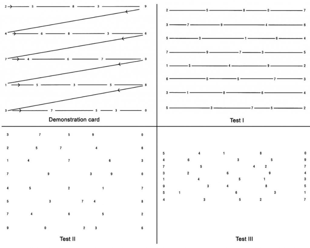
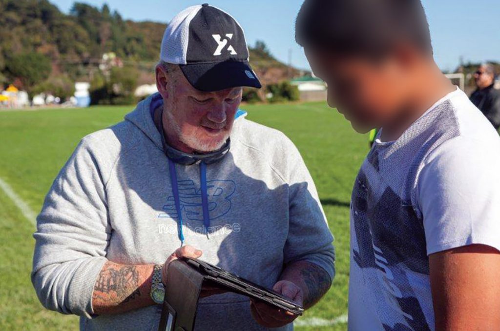
The King–Devick test (KDT) was developed as an indicator of saccadic performance as it relates to reading ability [4]. The KDT requires a player to read a series of single digit numbers displayed in the form of a “card” on a 10.1 inch android tablet, on a full-sized iPad, or from a standardised cardboard flipchart, taking 1-2 minutes on average. The test cards become progressively more difficult to read due to variability of spacing between the numbers. The time to read each card is recorded, and a “total time” is calculated. The total errors in reading the numbers are also recorded. A baseline test score for “total time” is recorded as the fastest time without errors that a player can read all three cards. After a potential injury either an increase in the errors made while reading or a slowing of reading speed are included in deciding if a player fails the test. Therefore, this is not just a test of saccadic eye movements, but also attention and language. The downside of the KDT is that it relies on the players having a baseline test score documented.
The KDT has been validated for use as a concussion screening tool. In MMA fighters post-fight KDT scores were significantly worse (higher) than pre-fight scores for participants who had sustained head trauma during the match [8]. Other studies have shown the ability of the KDT to detect concussion in hockey, lacrosse, football, basketball and rugby, with a sensitivity of 86% and a specificity of 90%. On average the time to perform the saccadic tests increased by 4.8 seconds from baseline in concussion [9].
In our own practice we have found the KDT to be a useful test to detect functional change in rugby players with documented head injuries. Furthermore, we found that stunt performers showed a significant change in KDT scores after falls from heights of 4 metres (in press).

Eye movement abnormalities

Cranial nerve palsies would not be expected in the setting of concussion or mTBI. An acute third, fourth or sixth nerve palsy would suggest either an intracranial haemorrhage or a pre-existing space occupying lesion. However, convergence insufficiency is well recognised following mTBI, with one study reporting 42% of athletes showing abnormal convergence one month after concussion [10]. The convergence or near triad is triggered by retinal disparity, and produces bilateral adduction of the eyes, miosis and accommodation of the crystalline lens. Therefore, it involves both afferent and efferent pathways. Convergence is tested, by measuring the “near-point” or the closest point to the face at which the patient can maintain binocular fusion. A change in the near point of convergence can be tested at the sidelines and compared to a baseline measure, but is not typically done until the player is back in a clinic setting.
Laboratory based tests
Visual tracking tests
Predictive visual tracking requires cerebellar coordination based on retinal inputs as well as higher visual processing, attention and working memory. Changes in visual tracking have been reported in patients with mild TBI [11]. Mild TBI patients displayed impaired target prediction with increases in eye position error. A circular tracking test, such as that used in the Sync Think (Boston, USA), has been shown to be a robust test to distinguish mild TBI from control patients [12]. However, other available devices such as RightEye (Maryland, USA) use a combination of circular, vertical and horizontal smooth pursuits and saccades in the testing protocols.
Visual evoked potentials
Visual evoked potential (VEP) are derived from changes in the electroencephalogram (EEG) measured over the occipital lobes in response to a visual stimulus. The waveform of the signal recorded can be quantified in terms of the size of the signal (amplitude) and the timing of the signal conduction along the visual pathways relative to the visual stimulus (latency). Latency changes have been shown to be significantly increased in those with mTBI compared with controls [13]. Further studies on VEPs have shown change in alpha rhythm attenuation in patients with mTBI and subjective attention deficits [14]. Further work is required to see if intra-subject changes in latency can be used to objectively detect concussion.
Optical coherence tomography
Mouse models of repetitive mTBI have shown a detectable thinning of the inner retina on spectral domain optical coherence tomography (OCT) [14]. There are no human studies published to date replicating these changes, but OCT may prove to be an in-vivo measure of cumulative concussive damage.
Treatment
Photophobia and visual discomfort when reading have been reported by mTBI patients. Using the Intuitive Colorimeter System, subjective improvements in visual comfort levels were found in 11/12 patients while wearing tinted lenses, however objective tests of reading parameters and VEP latency were not significantly altered [15]. Authors suggest these lenses could be used as an adjunct in managing post-concussion photosensitivity.
Convergence insufficiency has been shown to be more common in athletes with higher scores on symptom scales [10]. Treatment aimed at retraining convergence efforts and specialised reading exercises may therefore improve symptomatic issues (blurred vision, double vision) after concussion. The King-Devick Reading Acceleration Program is one example of a commercially available treatment protocol. Vestibular rehabilitation may also assist in improving visual function, by improving the vestibulo-ocular reflex and depth perception [4,5].
Conclusion
Concussion events occur with contact sports and may lead to subtle but cumulative damage in brain morphology and function [3]. Visually-based concussion tests are of value on the sidelines and in the clinic setting. However further research is still needed. Vision-based tests may prove to be the most reliable, portable and easiest to implement in our schools and in our amateur sporting teams. In the long-term these tests may be able to guide rehabilitation programmes, and allow us to assess when it is safe for a player to return to school, training, work and to the competition.
References
- Centers for Disease Control and Prevention. (2010). Rates of TBI-related Emergency Department Visits, Hospitalizations, and Deaths — United States, 2001–2010.
- McCrory P, Meeuwisse W, Aubry M et al. Consensus statement on concussion in sport – the 4th international conference on concussion in sport, held in Zurich, November 2012. J Sci and Med Sport 2013;16:178-189.
- Dimou S, Lagopoulos J. Toward objective markers of concussion in sport: a review of white matter and neurometabolic changes in the brain after sports-related concussion. J Neurotrauma. 2014;1;31(5):413-24.
- Ventura RE, Balcer L Galetta S, Rucker JC. Ocular motor assessment in concussion: current status and future directions. J Neurol Sci 2016;361:79-86.
- Ellis MJ, Leddy JJ, Willer B. Physiological, vestibulo-ocular and cervicogenic post-concussion disorders: an evidence based classi cation system with directions for treatment. Brain Inj, 2015;29(2):238-248
- King NS, Crawford S, Wenden et al. The Rivermead Post Concussion Symptoms Questionnaire: A measure of symptoms commonly experienced after head injury and its reliability. J. Neurol 1995;242(9):587–92.
- Galetta K, Barrett J, Allen M et al. The King-Devick test as a determinant of head trauma and concussion in boxers and MMA ghters. Neurology 2011 ; 76(17): 1456–1462.
- Galetta K, Lui M, Leong D et al. The King-Devick test of rapid number naming for concussion detection: a meta-analysis and systemic review of literature. Concussion 2015;10.2177.
- Pearce KL, Sufrinko A, Lau BC, et al. Near Point of Convergence After a Sport-Related Concussion: Measurement Reliability and Relationship to Neurocognitive Impairment and Symptoms. Am J Sports Med. 2015;43(12):3055-61.
- Barnes GR. Cognitive processes involved in smooth pursuit eye movements. Brain and Cognition, 2008; 68(3): 309-326.
- Suh M, Kolster R, Sarkar R et al. Defects in predictive smooth pursuit after mild traumatic brain injury. Neuroscience letters 2006;401(1-2):108-113.
- Chen XP, Tao LY, Chen AC. Electroencephalogram and evoked potential parameters examined in Chinese mild head injury patients for forensic medicine. Neurosci Bull. 2006;22(3):165-70.
- Yadav NK, Ciuffreda KJ. Objective assessment of visual attention in mild traumatic brain injury (mTBI) using visual-evoked potentials (VEP). Brain Inj, 2015;29(3):352-65.
- Tzekov R, Quezada A, Gautier M et al. Repetitive mild traumatic brain injury causes optic nerve and retinal damage in a mouse model. J Neuropathol Exp Neurol 2014;73(4):345-61.
- Fimreite V, Willeford KT, Ciuffreda KJ. Effect of chromatic filters on visual performance in individuals with mild traumatic brain injury (mTBI): A pilot study. J Optom 2016 May 30. pii: S1888-4296(16)30009-7.

