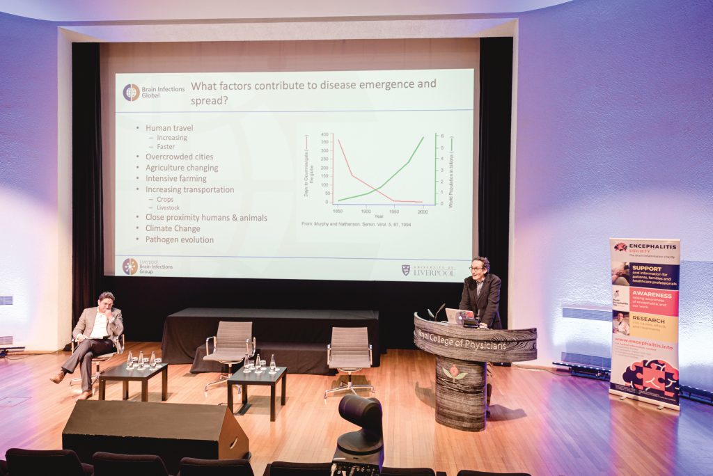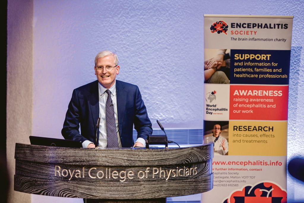
The 2020 Encephalitis Conference successfully took place during the 2020 COVID-19 pandemic. Despite these challenging circumstances the conference was delivered with a few key personnel present in-person and outside of this digitally to 257 delegates from 34 countries, welcoming researchers and healthcare professionals from around the world interested in a wide range of subjects related to encephalitis.
The Conference began with a session chaired by Dr Benedict Michael, Vice Chair of the Society’s Scientific Panel; Consultant Neurologist and Senior Clinician Scientist Fellow at the University of Liverpool, UK.
Dr Cecilia Zivelonghi, from the Department of Neurosciences, Biomedicine and Movement sciences at the University of Verona, Italy, gave a first presentation on SARS-CoV-2 infection. She discussed how COVID-19, a disease that mainly causes respiratory symptoms, also involved anosmia and ageusia, which led to the question of possible central nervous system (CNS) infection. They retrospectively analysed samples referred for antibody screening for SARS-COV-2 IgA and IgG testing, from March 1st 2020 to August 31st 2020. Among 339 patients referred for antibody testing, 23 showed either SARS-CoV-2 IgA and/or IgG (IgA n=9, IgG n=1, IgA and IgG n= 13). Among 21/23 available CSF, 4 were positive (IgG n=3, IgG and IgA n=1). Clinical features, available in all 23 cases, revealed encephalopathy (n=18) and seizures (n=8) as common manifestations and, in 4 cases, myelitis, predominantly with lower limbs weakness. 17/23 patients were systemically asymptomatic. Brain MRI showed FLAIR-T2 hyperintensities in 13/18 patients. EEG showed alterations including epileptic discharges (n=5) and/or generalized slowing (n=12). CSF pleocytosis (>5 cells/µL) was reported in 9/19 investigated cases. The clinical and radiological characteristics were compared with a group of 75 seronegative patients. Autoimmune neurology screening among seropositive cases revealed one patient with serum titin autoantibodies, one with limbic encephalitis and seizures had serum and CSF amphyphisin antibodies, and one presenting with acute disseminated encephalomyelitis had serum and CSF MOG antibodies. The incidence of SARS-CoV-2 IgG/IgA positivity was higher (7.8%, 18% when considering only patients with suspected encephalitis) than that reported in the Italian population (2.5%) and the observed clinical spectrum of disorders suggest that SARS-CoV2 could trigger inflammatory CNS processes, usually not associated with well-known autoantibodies.
Dr Yvette Crijnen, from the Erasmus University Medical Center, Rotterdam, presented a study about the autoimmune aetiology of new onset status epilepticus. Only patients without a known cause were included. Status epilepticus is commonly observed in autoimmune encephalitis, and, in this case, it is best treated with early immunotherapy. Dr Crijnen presented 50 patients with new onset status epilepticus, of whom 38% had a definite or probable autoimmune aetiology. Compared to patients without an autoimmune aetiology, patients with a definite or probable autoimmune cause were younger on average. They more frequently had a super-refractory status epilepticus, a tumour, behavioural changes preceding status epilepticus, abnormal mesiotemporal hyperintensities on MRI scans, as well as increased levels of white blood cells in the cerebrospinal fluid (CSF). A strong majority of the patients with a definite or probable autoimmune cause became seizure free after one year, especially those with antibodies against extracellular antigens. Their study demonstrated that status epilepticus with unknown aetiology frequently has an autoimmune aetiology. Neuronal antibody testing, MRI and CSF assessment can help identify such aetiology in patients with status epilepticus.
Dr Laura Bricio-Moreno, from the Massachusetts General Hospital and Harvard Medical School in Boston, USA, presented on the role of CXCL1 in viral encephalitis, a peptide produced by astrocytes and neurons in response to herpes simplex virus-1 infection, a disease associated with increased CSF neutrophils and inflammatory cytokines. Although treatment has shown a reduction in mortality, the morbidity of such often remains. Dr Bricio-Moreno showed that mice models with HSV encephalitis also had a majority of neurological morbidities, involving blood-brain-barrier (BBB) permeability and increased infiltration of neutrophils and inflammatory monocytes into the brain. Increased presence of CXCL1 was observed, but when deficient in CXCL1-associated receptor CXCR2, mice had reduced clinical severity, a higher rate of survival, reduced recruitment of neutrophils, decreased BBB leakage and viral morbidity. In addition, when neutrophil were depleted in WT mice, there was increased survival and reduced clinical severity, suggesting that neutrophils mediate HSV outcome and morbidity. This work shows that the CXCL1-CXCR2 axis could be a therapeutic target for limiting the morbidity linked to over-exuberant immune response in HSV-1 encephalitis. The potential of corticosteroids to help neutrophil migration was discussed.
Professor Tom Solomon, President of the Encephalitis Society and Professor of Neurology at the University of Liverpool, UK, concluded the first morning session with a presidential address entitled “Encephalitis Research in the Next 25 Years”. Prof Solomon highlighted the importance of surveillance in the identification of infectious and autoimmune encephalitis, but also in those with unknown aetiology. International spread of animal or arthropod-borne viruses has increased in the past centuries in part due to the amount and speed of human travel, overcrowded cities, agricultural progress, climate change, and pathogen evolution. Creation of zoonotic research hubs and development of machine-learning algorithms to predict viral phenomena, newer approaches to diagnosis, and more research on viral co-infections are emerging. Treatment development is also guided by the number of clinical trials which needs to be increased, especially in rare conditions. Furthermore, vaccines are being developed and more widely used with the current pandemic demonstrating how fast things can happen. The need to anticipate new pandemics in Neuroscience, the importance of raising awareness of the excellent risk-benefit ratio of vaccines, the potential of neuroprotective treatments for elderly populations and the need to improve access to basic clinical protocols were discussed.

The second morning session was chaired by Dr Jessica Fish, clinical psychologist and lecturer at the Institute of Health & Wellbeing, University of Glasgow, UK.
Dr Mette Scheller Nissen, from Odense University Hospital, Denmark, presented on 55 patients with anti-NMDAR encephalitis. The majority had a definite diagnosis of autoimmune anti-NMDAR encephalitis, and they could be subdivided into three categories, pure-, post-HSE or NMDAR encephalitis alongside other comorbidities (paraneoplastic and demyelinating). Memory impairment and sleep disturbances were rare in children, and abnormal movements were rare in adolescents. Cognitive decline, behavioural and psychiatric symptoms were relatively common in all age groups. The majority of the cases had abnormal CSF and EEG observations, and MRI abnormalities were found in approximately half of patients. PET revealed a majority of abnormal scans, mostly occipital and fronto-temporal, in almost half of patients with a normal MRI. Almost all of them received first line therapy and maintenance treatment, with only a quarter of patients receiving second line treatment. In an average follow up of two years and two months most patients with pure NMDAR encephalitis had a good clinical outcome, with much less among those with post-herpes simplex or other types. Awareness in paediatric patients and fast diagnosis and treatment remains important.
Dr Greta Wood, from Royal Liverpool University Hospital, UK, introduced a retrospective study on the predictors of seizures in encephalitis. Seizures affect large proportions of patients, although its effects and risk factors are unclear. Dr Wood presented a cohort of 203 patients from 24 hospitals across England, in which 121 had one or multiple episodes of seizures, or status epilepticus. The three most common aetiologies were infectious (including HSV), autoimmune (including encephalitis associated antibodies), or unknown. Most patients had seizures, focal and/or generalised: they were frequently multiple but there was also status epilepticus. Seizures were significantly associated with antibody or HSV related aetiologies. These were also associated with fever, reduced Glasgow Coma Scale (GCS, measuring levels of consciousness) on admission, and a worse clinical outcome. The aetiology, reduced presence of focal neurology signs, and lower GCS were the key predictors of seizures both during the first stage of the illness and in the inpatient period. This confirmed past research on correlates of seizures in encephalitis: the data highlighted the need to identify potential outcomes and anticipate their consequences. The discussion mentioned the lack of associations with MRI abnormalities and the possible findings of quantitative analyses, as well as the need to identify underlying mechanisms.
Vasundharaa S Nair, a PhD researcher at the National Institute of Mental Health and Neurosciences in Bangalore, India presented factors that limited or facilitated reception of care in India, for persons with Acute Brain Infections. Patients had diverse social and religious backgrounds, and most participants had a low socio-economic status. Ms Nair exposed two types of pathways that led to access to neurological services care. Most patients followed the medical model that directly went through general practitioners referring to the services. A minority went through traditional methods: relatives would first refer to a traditional healer, with rituals lasting days against a perceived curse or a divine punishment. Some were also advised to meet priests in churches due to sin or possession, who then would refer them to a physician. The latter would then give referral to neurological services. The presentation also showed the costly impact of health care requests, requirement of funding schemes, concerning the need for psychoeducation, work support or follow-up therapies. Lack of awareness, cultural practices, misinformation, unaffordability or unavailability of care, lack of adequate policy planning were the main barriers to treatment. Facilitators were the understanding of the medical model by religious referents, adequate awareness and acknowledgement of the disease, and accessibility to physicians and healthcare services.
Dr Frederik Bartels, from the Charité University Hospital at Berlin presented a study describing longitudinal changes in brain volumes of patients with anti-NMDA receptor encephalitis. Considering past reports of cases with long-term atrophy, Bartels presented quantitative analyses of MRI scans that were run over a median follow-up period of three years in a cohort of patients from the German Network for Research on Autoimmune Encephalitis. Around half of the cohort presented with abnormal MRI scans. The analyses allowed to test differences in brain volumes from the first to the last MRI. They showed that the annualised percentage of brain volume decreased over time while ventricular volumes increased. More specifically, white matter, but not grey matter, was subject to a significant decrease in the following scans. The volume reduction rate exceeded the pathological cut-off values that had been defined in multiple sclerosis with a similar age. High heterogeneity in volume trajectories and severe atrophy were also observed in individual evaluations. This led to the conclusion that brain atrophy was present in anti-NMDAR encephalitis, although with high variability in individual brains, specifically driven by white matter loss.
The morning sessions ended with the first Keynote lecture by Professor Carsten Finke, from the Berlin School of Mind and Brain and Charité University Hospital, Berlin. Prof. Finke focused on the information about the cognitive deficits in the years following anti-NMDA receptor encephalitis. Although many patients have a good neurological outcome using the modified Rankin scale, the discrepancy with persisting cognitive deficits was pointed out. One explanation for this is the impact of NMDA receptors in the hippocampus, a key region for memory processing. Mostly executive functions and memory deficits are found in long-term outcome. Predictors of these deficits included delayed treatment, higher disease severity and older age. Routine MRI reported relatively rare abnormalities but was found to be a predictor of long-term deficits. Functional MRI may be more sensitive: studies show abnormal spontaneous brain activities and reduced connectivity between regions, including the hippocampus, but also large networks correlated to cognitive performance. These differences also changed over time in dynamic connectivity analyses. Volumetric analyses of MRI scans also demonstrate volume and shape alterations of the hippocampus and white matter tracts, both correlating with cognitive performance. MRI studies are also relevant in children where much higher abnormalities and alterations were found in long-term brain development. Potential functional neuroplastic improvement in younger patients was discussed.
After lunch Dr Nicholas Davies, Chair of the Encephalitis Society Scientific Panel and Consultant Neurologist at Chelsea and Westminster, Charing Cross and the Royal Marsden Hospitals, London, UK, chaired the third session.

Dr Priya Thomas, from the National Institute of Mental Health and Neurosciences, in Bangalore, India, presented an exploratory study aiming at identifying the consequences of encephalitis in young adults and their families. Patients in South India who presented in a neurology department in a tertiary referral care centre were interviewed to collect information about how remission from the disease was managed by families and healthcare services with limited resources. How the illness changed their lives and relationships, the need for accurate information about what to expect, and the need for care support of families were recurrent themes. In the weeks following discharge from acute care, patients experienced a range of symptoms including headache, fatigue, seizures, motor and language difficulties, or memory loss. Dr Thomas concluded that it is important to understand and identify the consequences of the disease and its varying aetiologies, so that better health care and long-term support can be provided in these families and communities. The opportunity for these findings to impact institutional and regional policies in India was also raised during the discussion.
The next presentation was led by Dr Anna EM Bastiaansen, from the Erasmus University Medical Center, Rotterdam. Dr Bastiaansen presented an observational nationwide study of a Dutch cohort diagnosed with antiNMDAR encephalitis. The cell-based assays and immunochemical analyses that were run proved to be more sensitive to the diagnosis of the disease when dealing with CSF cells rather than blood serum cells: this was an indication that CSF tests should be recommended when suspecting anti-NMDA receptor encephalitis. Patients with an onset of the disease after 45 years had less frequently behavioural symptoms, seizures and seropositivity. Furthermore, they had a higher rate of delayed improvement (in coming back to an independent lifestyle), poor outcome after 1 year, and death. Common association of the disease with cancerous carcinoma was found in elderly patients. It also showed that second line immunotherapy led to recovery in a median of 61 days. This demonstrated that late onset of anti-NMDAR encephalitis is not as uncommon as previously thought and can lead to a worse outcome and delayed remission.
The second keynote lecture was given by Dr Benedict Michael, Vice Chair of the Society’s Scientific Panel, Senior Clinician Scientist Fellow at the NIHR Liverpool and Honorary Consultant Neurologist at the Walton Centre, Liverpool, UK. Dr Michael presented how the SARS-Cov2 virus affected the brain, the evidence known to date and how it can be addressed. As encephalitis involves infectious and auto-immune inflammation with direct and consequent phenomena, COVID-19 could be the cause of associated neurological disorders. Although biologically plausible, the evidence is still debated as encephalitis was found in low proportions of patients, with unspecific effects. However, there may be a neglect of symptoms resulting from cerebrovascular events like headaches and anosmia, as well as data that clinicians do not have the time to collect. Neuropsychiatric and cerebrovascular events were found in several patients with COVID-19 including delirium and inflammation in the nervous system. Cases of encephalopathy have also been found with MRI, but findings may be limited due to the difficulty to scan ventilated patients. Dr Michael presented the COVIDClinical Neuroscience Study that is currently investigating neurological complications, their mechanism, the role of biomarkers, and how to prevent long-term disabilities. Unresolved questions remain about the possible continuum of encephalitides across patients, including those that may have been unidentified.
Josephine Heine, a cognitive neuroscientist and PhD researcher at the department of Neurology at Charité University Clinic in Berlin presented a study that looked at health-related quality of life (HRQoL) in recovering patients with anti-NMDA receptor encephalitis. The study showed that patients with antiNMDAR encephalitis reported a lower HRQoL compared to the norm, despite good physical outcomes. Ms Heine exposed perceived burdens years after the onset of the disease, including seizures, fatigue and sleep problems, cognitive symptoms, and affective & behavioural symptoms. Additionally, higher prevalence of depression and anxiety, and reduced sleep quality was correlated negatively with HRQoL. Among subjective burdens, persisting seizures had the worst effect on HRQoL. Factors that correlated with higher HRQoL were greater day-to-day independence, fewer depressive symptoms, higher self-efficacy and higher satisfaction with one’s own memory abilities. The latter was higher than the population norm, even though actual lower memory performance was found, which might reflect a bias resulting from remission. Less frequent use of negative stress coping strategies also contributed to a better HRQoL: this could benefit from behavioural intervention. The study therefore highlighted the longterm contribution of underreported factors that affect quality of life and may require further support during recovery from antiNMDAR encephalitis.
This first afternoon session concluded with research funded by the Encephalitis Society. The first study was presented by Dr Aline de Moura Brasil Matos, neurologist and PhD researcher from the Tropical Medicine Institute at the University of São Paulo, Brazil. She gave a focus on demographic and clinical data obtained in patients with Chikungunya virus (CHIKV), followed in Brazil from 2017 to 2019. CHIKV is also thought to be an aetiological agent in encephalitis: the latter was detected in the majority of patients, among whom 37.5% died. The main mortality risk factors included hypertension, diabetes, required mechanical ventilation, seizures, acute kidney injuries, male gender and an age over 60. The relation of neurobiology data and population dynamics was discussed.
The second study was presented by Charly Billaud, a PhD student from the Institute of Health and Neurodevelopment at Aston University, Birmingham, UK. He presented behavioural and emotional profiles in a cohort of children with autoimmune encephalitis, which raised concerns about the difficulties faced among peers, conduct, emotions, prosocial behaviours, and hyperactivity. Overall, these difficulties were considered slightly raised in the general population norm but showed proportions of children with specifically high difficulties in these different areas, in a similar fashion as children with general neurology disorders. Behavioural analyses are to be used in a project involving neuroimaging-guided predictions that may help clinicians anticipate these future difficulties.
The last session was chaired by Ava Easton, CEO of the Encephalitis Society and Honorary Fellow, Dept. Clinical Infection, Microbiology and Immunology at the University of Liverpool. Dr Easton interviewed Brian Deer, an investigative journalist known for his inquiries into the drug industry, medical and social issues for The Sunday Times. He provided a reading from his new book “The doctor who fooled the world”, which explores disgraced doctor Andrew Wakefield’s controversial war against MMR vaccines. Deer exposed how clinical research was manipulated and conducted unethically, to the detriment of both the deceived public and the patients and their families, for the purpose of proving a link between the MMR vaccine and autism that in fact did not exist. The interview went into details about how Wakefield’s attempt to prove that vaccines caused neurodevelopmental issues had gone through grievous biases and research misconducts, as well as cover ups and costly legal battles. This was an example of deception and fraud that needs to be dealt with in biomedical research, not only on the part of Wakefield but also the system that allowed such research to happen.
Dr Ava Easton began to close the conference with a Call to Action relating to the Encephalitis Society’s situation going through the pandemic. She showed a video summarising what the Encephalitis Society has gone through during this pandemic year including how the charity has pivoted quickly and developed digitally for their beneficiaries. They have however lost significant fundraising events leading to income shortfalls, in direct contrast to a significant increased demand for help.
Dr Nicholas Davies and Dr Ava Easton presented the awards and prizes for best poster and best oral presentations:
Best poster for “Factors predicting patient quality of life after LGI1- antibody encephalitis” to Dr Sophie NM Binks, from the Oxford Autoimmune Neurology Group, at the Nuffield Department of Clinical Neurosciences of the University of Oxford, UK, (with colleagues M Veldsman, S Jacob, P Maddison, J Coebergh, S Michael, S Ramanathan, Easton A, M Scheller Nissen, M Blaabjerg, Leite M Isabel, D Okai, M Husain, SR Irani).
Best oral presentation for “Barriers and Facilitators in seeking care for Persons with Acute Brain Infections” to Vasundharaa S Nair from the Institute of Mental Health and Neurosciences, Banglalore, India.
The conference concluded with thanks to the conference sponsors and a closing call to action from from of Dr Ava Easton and Dr Nicholas Davies to get involved with World Encephalitis Day on 22nd February.
Encephalitis 2021 will be held at the Royal College of Physicians, London on 7th December 2021. You will be automatically notified if you are a professional member of the Society (membership is free) or you can find out more here: www.encephalitis.info/conference