On July 16th 2018, a neurosurgical update was organised in Aberdeen with a view to invite Scottish colleagues. This meeting was catered for the multidisciplinary team including neurosurgeons, electrophysiologists, anaesthetists, speech & language therapists, physicists and even a medical student (me!). More importantly, the event became truly special because of our keynote speaker Professor Atul Goel, a well renowned neurosurgeon from India.
Mr Mahmoud Kamel, Consultant Neurosurgeon at Aberdeen Royal Infirmary, opened the conference with a presentation titled “Endoscopic treatment of anterior skull base meningiomas. Is it worth the hassle?” In this he discussed the pros and cons of using an endoscopic approach vis-à-vis conventional transcranial microsurgical approach – specifically in cases of tuberculum sella meningiomas.
Our keynote speaker, Professor Atul Goel, addressed the “Planning of Complex Brain Tumours”. In this he shared his experiences with giant pituitary tumours and the challenges they bring. He presented various interesting cases he had seen over the years and the approach he took to managing those. These were very challenging to surgically resect, due to their sheer size and the invasiveness – especially when invading the cavernous sinus. However, he emphasised the fact that these tumours are confined to their dural boundaries and therefore understanding the anatomy helps massively.
Following on from this, Professor Goel spoke about the functions and importance of the cavernous sinus. He has discovered key concepts about the cavernous sinus, such as its importance in temperature regulation and its vital role in vision.
After this, Mr. Adnan Shaikh shared surgical management of a young patient with a dominant hemisphere AVM undergoing excision under awake craniotomy by Mr Pragnesh Bhatt, Consultant Neurosurgeon (and Convenor for the meeting). Dr Alan Forster, Consultant Clinical Neurophysiologist, and Penny Gravill, Lead Speech & Language Therapist, spoke about their experiences in awake craniotomies. They presented their involvement in initiating this service in Aberdeen and illustrated what their involvement was in this particular case. A full pre-operative language assessment, testing auditory and reading comprehension, was conducted. This was conveyed to the rest of the team to help plan and assess suitability for the procedure. Intra-operatively brain mapping and speech arrest were performed to make the surgery safe. The conclusion from this was that every member of the team had a valuable role and what makes such an operation a success was their coming together as a TEAM (Together Everyone Achieves More).
After a lunch kindly sponsored by Safe Orthopaedics, Mr Likhith Alakandy, a neurosurgeon at Institute of Neurosciences, Queen Elizabeth University Hospital, Glasgow, outlined the anatomical variations of the vertebral artery at the craniovertebral junction (CVJ). It is important to understand, especially when planning surgeries involving C1 and C2.
Prof Goel then spoke about the technique he invented for atlantoaxial fixation. He described how he used lateral mass screws on C1 and pedicle screws on C2 to achieve fixation, which addressed a key problem of instability of the joint that required stabilisation. He also noted that through his method, the anatomical alignment of C1 and C2 was not required and found they had achieved 100% fusion in patients. He also spoke about the possible injury to the vertebral artery due to its course and referred back to what Mr Alakandy said, that anatomical knowledge and understanding is vital here.
Mr James Walkden, Consultant Neurosurgeon at Aberdeen Royal Infirmary, spoke on behalf of the department about their experience using the O-Arm. This was the first hospital in Scotland to use the O-Arm. The O-Arm is 3D computer assisted navigation based on CT imaging. It allows for multiple images to be taken which can be used to generate a 3D model. He noted that this imaging allowed for better accuracy for insertion of implants at the cranio-cervical junction as well as upper thoracic spine where routine fluoroscopy has some limitations. It was also highlighted that O-Arm is especially useful for training.
After a short break, Dr Brian Morrisey, Radiology Registrar at Aberdeen Royal Infirmary, gave an illuminating talk titled “Diagnostic uncertainty in High Grade Glioma: our experience learning from tumour chameleons”. He explained that brain tumours could mimic different diseases which are common. The tumour is said to be a chameleon since it can make itself look like a different pathology. He then presented a case series, in which 13 cases were identified via the MDT over the past two years. The most common tumour chameleons were ischaemic and haemorrhagic stroke, followed by encephalitis. Out of these, the initial presumed aetiology was an ischaemic infarct in five of the cases, while in another three it was a haemorrhage. Following histopathology it was found that seven of the 13 were GBMs. It was concluded that GBM misdiagnosis can occur and that encouragement of early repeat imaging may be able to catch these chameleons promptly.
To finish off the day, there was a presentation by Mr Aimun Jamjoom, an ST4 Neurosurgical Registrar, on behalf of the British Neurosurgical Trainee Research Collaborative (BNTRC). He spoke about a research collaborative which explored external ventricular drainage. They found that the median time to infection for plain catheters was 8 days. In silver impregnated catheters it was 7 days and in antibiotic impregnated ones it was 11 days. Mr Michael Poon, Registrar from Edinburgh also spoke on behalf of the BNTRC, presenting a study about Chronic Subdural Haematomas. He concluded that the preferred method of treatment was burr hole drainage with a postoperative closed drainage system.
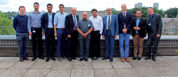
To end, a group photograph was taken, and the attendees were able to mingle with each other discussing future projects and collaborations.