
The Stroke Advanced Neuroimaging day was held in the heart of London at the Institute of Neurology. The Institute is located next to, and associated with, the National Hospital for Neurology and Neurosurgery, the largest dedicated centre for neurology and neurosurgery in the UK. This course aimed to deliver a focused review of the most commonly used imaging modalities in stroke and discuss how they are used to assess and evaluate brain infarct and haemorrhage. Within this context, there was an update on neuroanatomy as applied to neuroimaging, introductions to the application of mechanical thrombectomy and the imaging of stroke recovery.
We began the day with Professor David Werring giving a review of the clinical stroke syndromes in Applied Neuroanatomy for Stroke. Prof Werring’s concise presentation made a complex topic simple, as he stressed the importance of stroke syndrome localisation. He emphasised the usefulness of differentiating between the cortical and subcortical syndromes, both to provide clues on the aetiology of the stroke and also, crucially, to help predict progression over the acute period and determine optimal place of care.
Next, Professor Rolf Jager took us on a comprehensive, whistlestop journey through the Anatomy of the Cerebral Circulation. We were whisked up the carotids and through the arterial cortical territories, coursing around the Circle of Willis to the borderzone areas and the perforators before finishing in the venous system. Particularly interesting was a detour into the embryology of the cerebral circulation, including the genetically determined anatomical variants of the vessels. One striking example was that of an absent A1 segment of the Circle of Willis, resulting in an incomplete circuit, prevalent in different populations worldwide and conveying an increased lifetime stroke risk.
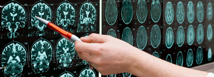
Next up, Professor Xavier Golay took us through his Introduction to Imaging for Stroke. We were taken through the inner workings of the stroke imaging modalities (CT and MRI) with interest kept high with memorable and absorbing facts. It was interesting to hear that CT sensors rotate at 100 times per minute, and that MRI measures the “spin” of nuclei but that only those nuclei with an odd number of particles can spin. His comprehensive diagrams helped non-specialists understand this difficult topic. We enjoyed being taken back to basic principles to understand the rationale for differing MRI sequences as applied to stroke medicine, including different types of MRI contrast such as PD, T1, T2, T2*, DWI, MRA, PWI and SWI.
Following this, Prof Jager returned to the lectern to present Neuroimaging 1: Ischaemic Stroke. We were taken through the pathophysiology within the acute timeframe of an ischaemic stroke. Prof Jager used example images at every stage to illustrate the transition from early to late ischaemic changes, later introducing the Alberta Stroke Programme Early CT Score (ASPECTS) used in CT in MCA stroke. We learned about clinical uses of different MRI sequences in stroke and heard about ongoing research using MR in wake-up stroke. Prof Jager also spoke about uses of neuroimaging in subacute stroke, in particular in elucidating aetiology and thereby, appropriate secondary prevention.
Next was Neuroimaging 2: Haemorrhagic stroke, presented by Prof Werring. Prof Werring took a stepwise approach to this topic starting with the aetiology of haemorrhagic stroke, taking us through the most common causes, including hypertension and cerebral amyloid angiopathy through the macrovascular causes including AVM and aneurysm rupture, and into rarer causes such as inflammatory conditions. We were then taken through practical guidelines in the investigation of the cause of haemorrhage, AHA recommendations on imaging and the uses of Digital Subtraction Angiography.
A lively panel discussion followed, chaired by Prof Werring and Dr Sumanjit Gill. The audience included UK-based and international stroke practitioners, and questions ranged from the controversies surrounding use of anticoagulation in patients with haemorrhagic stroke and concurrent atrial fibrillation to access to CT-angiogram reports in district general hospitals.
After lunch we returned to a fascinating lecture by Dr Peter Cowley on Endovascular Treatment. Dr Cowley took us through the landmark trials (Mr CLEAN, EXTEND -1A, ESCAPE, SWIFT-PRIME, REVASCAT) illustrating the different devices in use. A particular highlight was a video reconstruction of the device in action. This part of the day led nicely to the interactive cases chaired by Dr Cowley.
Three case presentations from stroke trainees followed, including cases such as successful thrombectomy in a palliative cancer patient. The cases stimulated further lively discussion from the floor.
This portion of the day was followed by a very interesting lecture on Imaging Stroke Recovery by Dr Tom Hope. He detailed his work using the PLORAS database (“Predicting Language Outcomes and Recovery After Stroke”) which has over 750 stroke patients and links their scores in detailed language and cognitive assessment with brain lesions on T1 MRI. He discussed three fascinating studies that used this database looking at predicting longitudinal change in chronic aphasia patients and prognoses in complex populations (including bilingual patients). Dr Hope’s work provides some context to taking neuroimaging beyond the acute stroke into prognosis prediction and potentially into treatment response.
The course was extremely informative and well-presented, with engaging and knowledgeable speakers. The course materials were easily accessible and will be a useful reference tool and a comprehensive reading list was provided for further study. I think the lectures would be hard to follow without a medical background but I highly recommend this course to those interested in gaining depth in their understanding about neuroimaging as applied to stroke medicine.