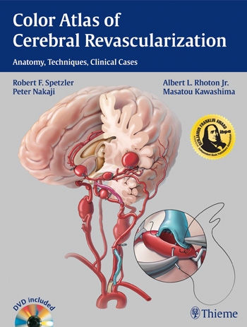
This formidable text represents a distillation of neurosurgical anastomotic technique to ameliorate (or prophylactically prevent) cerebral ischaemia, encompassing various types of vascular pathology. While describing a discipline that is being progressively supplanted by endovascular methods, the volume accentuates the need for surgical expertise in very particular situations. The distinguished authors have collected contemporary examples of their practice, the majority from the Barrow Institute in Phoenix, and have complemented their text with detailed photographs, line drawings and anatomical dissections. In this way each specific revascularisation takes place before the reader’s eyes in a sequential but simple fashion, highlighting correspondence with named structures at critical moments of the operation. As an additional and welcome adjunct, there is an excellent DVD focusing upon key cases, and this is invaluable for understanding the real-time theatre pressures that will always exist.
The book makes no attempt to revisit the complexities of diagnoses, indications for, and outcome measures relating to each procedure, and anyone expecting a breakdown of longer term complications (e.g. cerebral hyperperfusion syndrome, etc.) will be disappointed. Nevertheless, by means of brief vignettes of patients’ condition before their operation, the reader gradually gains a better grasp of alternative strategies that may be relevant in his or her practice. However this is a work principally concerned with techniques and technical advances. A significant proportion of the illustrated examples are clearly very demanding, while still being pragmatic in the correct setting. Accordingly it is likely that this book will really appeal to established vascular surgeons wishing to develop their repertoire (rather than junior trainees), although there is much that all could learn from the detail offered.
There are 16 chapters addressing the different bypass procedures, pertinent to both low-flow and high-flow revascularisation, with suitable emphasis upon the more common techniques. While the superficial temporal artery to middle cerebral artery section is very comprehensive (including double-barrel grafts and a large group of different aetiologies), there is also a separate chapter devoted to Bonnet bypass, and even facial – vertebral artery bypass. Everything from saphenous vein bridging grafts to complex skull base tumour resection with high-flow bypass is covered, and one can appreciate the common characteristics of the treatments despite quite varied pathologies. What soon becomes obvious is that, although standard microsurgical methodology will be familiar to most neurosurgeons, the sheer imaginative scope for alternative grafting that has arisen in the authors’ unit(s) is inspiring. Furthermore, they are not afraid to demonstrate their means of dealing with inevitable complications (such as vessel tears), and this realistic interpretation of emergency options is indeed refreshing.
UK neurosurgeons might look enviously upon the ease of intraoperative angiography within this case selection. While some specific types of instrumentation (e.g. the patented microsuction system, etc.) may be regarded as unnecessary, it is worth remembering that such approaches have evolved over years in an outcome-proven institute to maximise the chances of technical success; they should not be underestimated! Overall the authors and their support staff are to be congratulated on an excellent piece of work; I suspect that the majority of my Vascular Neurosurgery colleagues will rapidly add it to their library.
ACNR 2013;13:5:25

