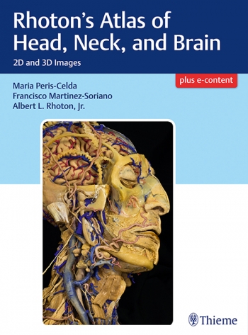
I first heard the name Rhoton when assisting in an operation to secure a ruptured brain aneurysm as a junior surgical registrar. The instruments which bear his name are used for microsurgical techniques in neurosurgery, helping to carefully dissect the brain to its deepest structures and secrets. After this operation, I decided neurosurgery was the career I wanted to follow, and Rhoton was a name I have heard a lot more of since.
Known to some as the father of microscopic neurosurgery, Rhoton’s reputation as an anatomist, surgeon and teacher became more apparent when I was learning more about the brain. This book encompasses not only the art and scientific detail of the dissections Rhoton was famous for, but also the way in which they are presented, conducive to learning anatomy, and sharing this knowledge with others.
In 624 figures arranged in 28 sections over four parts Dr. Peris-Celda and Prof. Martinez-Soriano lay out the head, neck and brain in stunning detail, with meticulous labelling by Prof. Valverde and Prof. Martí.
Part one covers the osteolology of the head and neck. One can spend many hours searching for well annotated plates such as these. Holding a 3D model of a skull in your hand whilst examining the images can be recommended, to fully appreciate the grooves, foramina and prominences.
The face and neck is covered in part two. The nerves, blood supply and muscles which give humans expression are beautifully dissected and displayed layer by layer. This helps establish a better understanding of the relationship between these structures, which leads to their function, but also when learning how to turn flaps and perform craniotomies.
In part three, the ear, nose, pharynx, larynx and orbits are shown. These are important parts of anatomy for neurosurgical trainees to be familiar with. Minimally invasive approaches to the skull base, such as the trans orbital, are becoming the mainstay of some practice. Although some of this anatomy is daunting to a junior trainee, the way that the authors lay out the dissections help digest the information and understand the function of their components.
Part four goes into intricate detail of the brain. These dissections help visualise the approaches used to different areas of the brain, and tie in the deficit which would be experienced in disease. Understanding of neuroanatomy in this detail enables the most complex of neurosurgical operations to be undertaken. Dissections rather than illustrations or diagrammatic figures serve this purpose to a greater extent when laid out well, as they are in this volume.
The book is large and heavy, reminiscent of coffee table hardbacks, and is not something to be carried round on a day to day basis. The price tag is equally hefty, more suitable for a departmental purchase than pocket money. That being said, it is a pleasure to use and would be a great addition to any neurosurgeon’s library. Perhaps the icing on the cake is that the plates are also available online. With the use of the 3D glasses provided, the dissections come to life. The view down the glasses is very similar to that of the operative microscope and helps further appreciate the relationship of the structures shown.
This book serves as a worthy tribute to the work of Rhoton, laying out beautifully dissections which help impart his skills and knowledge to the next generation of surgeons, and will help them to do the same to the following generation.

