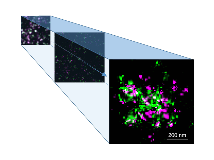
Direct images of a vital brain receptor, 20,000 times smaller than a human hair, have been captured for the first time as part of a world-first study that could allow scientists to unlock more information about neurodegenerative disorders like Alzheimer’s and Parkinson’s.
The international study, led by scientists at the University of Birmingham in collaboration with the University of Würzburg, examined G protein-coupled receptors (GPCRs), specifically the metabotropic glutamate receptor (mGluR4), which mediates the effects of the brain’s main neurotransmitter, in an attempt to establish how the receptors are organised within the brain. While previous research had investigated other types of receptors, very little was known about the organisation of metabotropic glutamate receptors and other GPCRs in the brain.

GPCRs play a fundamental role in regulating virtually all physiological processes in the body, enabling cells to sense external stimuli and communicate with each other. Because of this, they are the targets of at least one third of all drugs on the market. When cells are unable to communicate effectively, conditions such as Parkinson’s, diabetes and heart failure can develop. Understanding how these receptors work under normal conditions is key to the development of effective treatments.
By using an innovative super-resolution microscopy method known as dSTORM this latest study is the first to produce images of mGluR4 receptors in the brain with a resolution of approximately 10 nm, i.e. at least 20-times better than with conventional microscopy. The nanoscale images reveal that the receptors are not randomly located within synapses as previously assumed, but instead are located in highly organised so-called ‘nano-domains’, where they sit very close to the molecules that they regulate. This, researchers suggest, could mean that instead of sending messages to those molecules indirectly, the receptors may physically interact with them to regulate neural communication.
Davide Calebiro, Professor of Molecular Endocrinology at the University of Birmingham and Wellcome Trust Senior Research Fellow, who is lead researcher on the study, said:
By understanding how receptors such as mGluR4 work, we hope to be able to design innovative drugs capable of modulating their function. Our findings could help better understand neurological and neurodegenerative disorders and develop new treatments for diseases such as Parkinson’s or Alzheimer’s.
We hope this research could pave the way to further studies aimed at explaining the functional consequences of the high spatial organisation of synaptic GPCRs we have uncovered. In addition, it will enable other groups to apply similar methods to investigate the nanoscale organisation of molecules involved in other fundamental biological processes.
The next stage of the research is to study the movement of these receptors, as Professor Calebiro adds “So far we have obtained static pictures, but we suspect these structures to be highly dynamic. At our Centre of Membrane Proteins and Receptors (COMPARE), a joint venture with the University of Nottingham, we and other researchers are developing highly innovative methods to track single molecules in real time with unprecedented spatio-temporal resolution. We plan to use these methods to investigate the dynamics of receptors and other signalling molecules in neurons and other cells, such as heart cells.
The full paper ‘Super-resolution imaging reveals the nanoscale organisation of metabotropic glutamate receptors at presynaptic active zones’ was published in Science Advances.