Interview by GEMMA CUMMINS | Portrait by Paul Cash
A career highlight Realising, about 15 years after being appointed a Consultant Neurologist at Addenbrooke’s and five years after being appointed Clinical Dean in the University of Cambridge, that there was no place I would rather be in my career. I was a clinician working in a world class research centre, surrounded by the stimulation of young research students and clinical students and yet still spending much of my time in direct contact with patients.
Biggest regret That I probably never will write that novel, otherwise I refer readers to Edith Piaf.
Inspiring mentor Michael Harrison (at the Middlesex hospital), probably the best Neurologist of his generation and certainly the nicest, he inspired me in many ways personally and neurologically.
Most memorable patient There have been many, memorable for different reasons. One lasting impression was left by a patient with metastatic cancer I met as a clinical student. He said “I hope you’re not going to tell me I only have months to live, because some idiot of a doctor told me that 4 years ago!” This taught me never to specify any person’s lifespan…doctors are usually wrong when they try to guess how long someone has to live.
If I hadn’t been a neurologist When I was 10 years old, admiring my father, I wanted to be a fighter pilot. At school I was mainly interested in literature and art but good at biology, so medicine seemed to be a good practical choice. At various stages as an undergraduate I wanted to be a Psychiatrist, a Paediatrician and General Physician (internist) in that order (I never saw myself as a Surgeon). I only decided to commit to being a neurologist after nearly five years working post qualification in various medical specialties including two years as a general medical registrar. Already having an intellectual interest in the brain I realised that in neurology I would be able to practice the clinical method in its ultimately satisfying form, now aided by increasingly sophisticated neuroscience and imaging. Remember I started as a medical undergraduate the year levodopa was introduced as a treatment for Parkinson’s disease and qualified in the year CT imaging of the brain was introduced into clinical practice.
I now see that if I hadn’t become a neurologist I would have been very disappointed with my life (unless I had written that novel).
Hidden talent Having so few I prefer not to hide any talent I think I have!
Advice to budding Charcots (ie trainee neurologists) Never let someone else direct your career, believe in yourself but check your ambitions against reality. Try to end up doing something you are good at rather than something you wish you were good at. Disappointment in careers arises from a mismatch in a person’s ambition and his/her ability to achieve it. Take advice from multiple sources and then see what these different sources have in common. Always consider whether someone is advising you for their own benefit or yours. There is a lot of luck involved in the evolution of someone’s career but luck favours the prepared.
3 most important qualities in a neurologist He or she must be an empathetic physician. You must combine your knowledge of the science of disease and your clinical skills at identifying it with compassion for the individual, treating the patient and not the disease. Apply the scientific method to understand the disease and with this use human compassion to manage the patient’s illness. Try to be the neurologist you would like to consult.
Next frontier in neuroscience is… If I predict this I will be wrong. However I suspect it will be in the territory furthest from our understanding now, which is the frontier between the brain and the mind (and whether this is a discoverable border after all).
Guilty pleasure Eating too well. I don’t feel guilty about drinking too well and I’ve given up smoking long ago….as to other pleasures I feel no guilt.
Favourite tipple in favourite place A glass of American IPA anywhere with one or more of my grown up children, one a Neurologist, one a Clinical Psychologist and one on his way to becoming a Psychiatrist and all three, with their partners, my closest friends and the parents of my (soon to be) five grandchildren.
Favourite quote Voltaire is reported to have said “The effective physician is one who successfully entertains the patient whilst nature effects a cure”. When I told this to a neurosurgical colleague he said “I suppose you will be saying that the effective surgeon is one who successfully entertains himself whilst nature effects the cure”.
Most embarrassing moment When, on my first ward round as a clinical student at Guy’s Hospital, I realised that the zip on my trouser fly had totally failed.
Most cherished possession (apart from family) My Bamboo handled “Queen Square” style tendon hammer, a chimeric object derived from multiple sources over thirty years. It does more than obtain reflexes, it is my totem. Generations of registrars have been trained to retrieve it from lost regions of the hospital.
Fireside read Novels by the new generation of Indian and British Indian authors such as Amulya Malladi, Vikram Chandra and Amitav Ghosh.
Painting I would like on my wallA good one that I had painted, another unrealistic ambition, failing that “Sainte-Victoire Dagï” by Cezanne or David Hockney’s “Woldgate Woods”.
Desert island playlist I would take a collection which reminded me of various stages in my growing up:
Harry Belafonte “The banana boat song (Day O).” My father would play this frequently whilst I was a child growing up in India, where he worked as a Tea Planter. My father was the greatest man I have ever known, my icon still.
Cliff Richard “Livin’ lovin’ doll.” This came out when I was nearly 11 and my older brother played it endlessly in the school holidays.
Charlie Parker “My old flame.” Charlie Parker was my musical obsession at 16 when I was wrestling with the choice of whether to do science A levels or English and History.
Beatles “Love me do.” This peaked in the British charts at the same time as my last holiday visit to my parents in India; it marked the beginning of the crazy 1960’s as far as I was concerned.
Rolling Stones “Get off of my cloud.” A reminder of my somewhat disorganised life as an undergraduate.
Credence Clearwater Revival “Down on the Corner” Would remind me of more time in the “wasted” part of my youth.
Cat Stevens “Morning has broken.” My long suffering wife was a Cat Stevens fan when we met and this was played at our wedding in 1973.
Mozart Bassoon Concerto in B flat major, K. 191/186. I played Mozart cassettes nearly continuously in the car when travelling around East Anglia in anticipation of applying for my consultant post involving clinics in Cambridge, Peterborough and Kings Lynn.
Glasgow Caledonian University (GCU) researchers, in partnership with the University of Newcastle and Newcastle-based SME Peacocks Medical Group, have been awarded significant funding for the design and manufacture of innovative foot orthotics using 3D-printing technologies.
The project has been granted a £77,000 Small Business Research Initiative (SBRI) Healthcare development contract. SBRI Healthcare is an NHS England initiative, championed by the newly formed Academic Health Science Networks to develop innovative products and services that address unmet health needs.
The funding was awarded following a call to address challenges in improving diagnosis, self-management and prevention of musculoskeletal disorders.
Disabling foot and ankle conditions affect approximately 200 million European citizens. Over 300 million per annum is spent treating many of these people with orthoses and splints, often relying on hand-crafted manufacturing techniques which are slow, costly and difficult to reproduce.
With an increasingly ageing population and a growing health burden in long-term conditions, the global market for custom foot orthoses continues to grow.
The GCU team, led by Dr Gordon Hendry and Professor Jim Woodburn, will work with Peacocks Medical Group and researchers from Newcastle University on the ‘FootFEMan’ project, which will utilise a computational engineering tool called finite element analysis to improve the functional design of orthotic devices for individual patients.
The improved personalised design will then be printed layer by layer using 3D-printing techniques developed previously in the team’s award-winning EU-funded project, A-FOOTPRINT.
Dr Hendry said “We are confident that we can successfully 3D print new orthotic insole devices. This project will now enable us to improve each orthotic tailored to the individual patient according to whatever foot problem they have.
“We will test the new products in controlled clinical studies here at GCU to see if we can improve foot function during walking and further lessen disabling foot symptoms.”
Professor Woodburn added: “GCU’s collaborative partnership with Peacocks will enable them to maintain and grow their market position as the leading SME developing innovative and knowledge-based orthotic products.”
If you, or a member of your family, had an acute and potentially serious neurological problem would you expect to have the benefit of a specialist neurological opinion on the day of your admission to hospital? The Association of British Neurologists (ABN) surveyed all UK Neurologists, who were asked to provide details of their local services, which generated data for 195 acute hospitals across the country. The result is the Acute Neurology Report, the first national survey of acute neurological services, which has just been published by the ABN and reveals that on average across the UK a neurological opinion within 24 hours is only available in just over half of acute hospitals. At worst, in District General Hospitals without a dedicated neurology service, a same-day neurological opinion is only available 30% of the time, on days when a ‘visiting Neurologist’ is present. The thirty-one Neuroscience Centres in the UK mostly meet the highest quality standards by providing daily neurology specialist review and CT and MRI available 24 hours a day. But there remain a disturbingly large number of hospitals with no neurology service at all, with the Northern region, Northern Ireland, the North West, Wales and the West of Scotland having the most sites with significant gaps in neurology services.
This mixed model of neurology service provision, a reflection of the way the NHS has developed around small local hospitals, District General Hospitals and specialist tertiary referral Neuroscience Centres, inevitably leaves gaps and means the experience of a patient with a neurological illness is determined by geographical location rather than clinical need. It is well established that neurological input leads to improved diagnosis and shorter hospital stays, and it is reasonable to conclude that patients with neurological disorders admitted to hospitals without resident neurologists will have significantly worse access to appropriate investigations and services, which is likely to impinge on the quality of their care.
The ABN has proposed a set of Quality Standards, which define appropriate acute neurology care, and can be summarised as follows:
There should be access to daily consultation by neurology specialists within 24 hours of admission (if necessary by telemedicine) with care in an appropriate inpatient setting depending on clinical need (including the option of transfer to a neuroscience centre, neurosurgery or intensive care).
Advice on the management of acute neurological emergencies should be available from a neurology specialist at all times
Urgent inpatient imaging (CT and MRI), where indicated, should be available
Lumbar Puncture should be available at all times.
Rapid access pathways should exist for referral from Emergency Departments and Acute Medical Units to neurology outpatient services
The readers of ACNR will no doubt find these standards simple and obvious and may be baffled that they are not part of every hospital’s acute care policy. But the lack of a specific commissioning strategy for neurology has led to inequities in care. Although neurological services have expanded greatly, it is a matter of concern that the provision of inpatient neurological care has been neglected compared to other specialties such as cardiology and gastroenterology, with increased access to outpatient neurological services over recent decades being driven mostly by waiting list targets. Many of the commissioning services and NICE quality standards are disease specific and as a result patients with undiagnosed neurological conditions can be neglected by this process.
A significant step in the right direction would be the adoption by commissioners of the ABN’s Quality Standards, so that neurological services can develop to meet the increasing burden of an ageing population.
Andrew Bateman, Rehab Editor (and Quality Improvement Fellow in the same cohort).
People in need of our services need staff who offer clinical expert knowledge, skills and passion backed up by experience and research Good clinical leadership of our services is self evidently important in ensuring the patient experience is as good as it can be.
There are currently in the UK National Health Service a number of leadership training schemes. In the East of England scheme which is entering a third year of operation, a recent Quality Improvement programme has brought together a wide range of clinicians and administrators to be “Quality Improvement Fellows”.i Fellows had the chance to benefit from mentors and training provided by staff at the King’s Fund. I heartily recommend colleagues to look for similar opportunities.ii
In one of the sessions of this training programme participants enjoyed a moment to reflect on the art of story-telling. Graphic designer Graham Ogilvie was present, drawing simple cartoons of the messages conveyed by participants who had been asked to turn their quality improvement projects into a story. Participants were encouraged to use the form of a story, to think about characterisation, heroes and villains, start and ending, and other ingredients. I considered that Gemma has done this in such a compassionate and warm way, clearly demonstrating the point of Dementia Friendly wards.
There is a broader point: as scientist-practitioners we need to ask ourselves how we should best translate findings from projects (service improvement, audits or research) into documents that motivate action. Have we shared our learning in a way that ensures widest possible benefit?
Sometimes translation of research can be done in ways that are very familiar to us all, and it is perhaps not a fairy story to imagine that Services can improve through leadership such as is provided by people like Gemma. Gemma’s story provides an illustration of this because we are readily drawn into narratives. I wonder if you find yourself thinking about what you could do differently as a result of reading this?
Perhaps a different style of article to those usually published in this journal, I was really interested to note how it has been possible to allude to a rich literature in a short space. But most of all, I found this story to make for a compelling read and I hope our regular readers do too.
John and Mary had been married for 52 years, they lived a happy and fulfilled life together, they had raised their family, John had worked hard as an engineer travelling around the country and now they were enjoying their retirement together. John was extremely active, he enjoyed spending time with his family, working in his shed completing DIY tasks and maintaining his vegetable filled beautiful garden.8,11 Over a period of time a dark fog began to envelop John. He would be part way fixing the tyre on his bike and he would forget what he needed to do next.7,8 He began to get muddled with what day it was and forget the word that he wanted to say in conversation with his grandchildren.7 John began to withdraw in himself, he felt invisible, he felt his family would talk about him as though he was no longer there, he lost his appetite, he refused to drink, he would wear the same clothes for days and would stare out onto his beautiful garden lost in his own world.4,7,8 The fog deepened, John began a downward spiral, falling deeper and deeper into the unknown.4,8,15 Eventually John found himself trapped in a dungeon, guarded by dragons and tied up in chains of stigma, his voice and identity lost.4,15 Scared and confused by his surroundings John looked for ways to escape but the dragons kept pulling him back.5 John tried to fight them, he kicked and hit, but the dragons overpowered him with potions that made him tired and sleepy.3
One day John woke from his sleep to find a beautifully weaved basket full of items that made up his identity – a photo of his and Mary’s wedding day, the boiler suit he loved to wear when outside working and his gardening book that he loved to flick through whilst having a cup of tea in the morning.2,8,10,11,15 As John picked each item from the basket a calmness descended upon him and before his eyes an angel appeared – a Dr Angel – who granted him three wishes.6,10,11
John’s first wish was to get his voice back – he no longer wanted to be invisible and he wished his thoughts and beliefs could be heard.3,8,11,13 His second was to be able to do the activities he previously loved to do, to have a role, and feel useful and needed.7,8 His third wish was to have the opportunity to go home to his lovely wife and familiar surroundings.
Suddenly the fog began to lift and the dungeon was no longer a dungeon, it had transformed into a dementia friendly ward.4 The environment was a large space with clearly visible signs to direct to the toilet. John was encouraged to walk around. He had access to a garden and fresh air.4,5,7,13,15 The dragons became health professionals that would call John by his name, listen to his wishes and act upon his request for support of his care needs.4,7,12 They communicated with John with an improved level of respect and were mindful that John may find complex sentences difficult to process.7,13,14 They were patient and understanding when John struggled to clearly express himself, but gave him the opportunity to have his say.2,7,12 John was enabled to complete his own personal care tasks and given support when needed.8,13 They encouraged him to dress in his own clothes and worked with John at a level he was able to understand. John became involved in ward activities. His skills in DIY were actively encouraged to help build the raised flower beds in the garden and care for the many herbs and flowers donated to the ward.3,5-7,10-13 Alongside the increase in his activity, John’s appetite began to return and when he woke one morning craving a bacon and egg sandwich, one was sought after.1,8 Mealtimes were encouraged to be social events where all sat at a brightly coloured table set for the gentlemen in the bay.5,11 Drinks were offered throughout the day and snacks of sandwiches, cakes or fruit were freely available to pick at when peckish.6,7
John and Mary were included in the discussions of his ongoing medical care and his voice was loud and clearly heard when planning his discharge from the ward.3,6 John was given the opportunity to go home, with 24 hour enabling support that reduced over a 21 day pathway. John and Mary were involved in the process of tailoring the care to enable and support John’s needs, re-establish his routine and set himself goals.3,7,9,13 Mary was given support in managing the times when John became frustrated with his slow recovery and difficulty in understanding what had happened (that it had seemed like he was living in a nightmare).3,14,15 For the 21 days the carer, Mary and John could seek advice and support over the phone, and home visits took place to refresh John’s goals and to see his functional improvement in his home environment.7,8 The carer encouraged John to engage in the activities he previously enjoyed.2 Things began to settle and one day into the second week Mary looked out of the kitchen window and began to smile at the sight of John and the carer caterpillar hunting around the vegetable garden, sneakily eating the blackberries until their tummies ached.2,8,10,13 The pathway provided education to Mary on the ways to reduce the risk of the dungeon and dragons returning again. By monitoring John’s medication compliance, nutritional and fluid intake, bowel and bladder habits as well as being aware of changes in John’s behaviour.3,4,7,14,15 Midway through the pathway a mental and physical health review in ambulatory care signed John off from the acute hospital and he was referred on for diagnosis, advice and support on dementia.14 John no longer felt invisible and alone. He began to realise that this was going to be a new stage in his and Mary’s journey together.2,7,8 For John the fog was still there, but it was lighter now and the sunshine began to break through.4,8
Bruton A, Lipp A, McKenzie G. Graduate foundation scheme with a focus on dignity and older adults. Nursing Management -2012;18(9):20-5.
Carr TJ, Hicks-Moore S, Montgomery P. What’s so big about the ‘little things’: A phenomenological inquiry into the meaning of spiritual care in dementia. Dementia 2011;10(3):399-414.
Clissett P, Porock D, Harwood RH, Gladman JRF. Experiences of family carers of older people with mental health problems in the acute general hospital: a qualitative study. Journal of Advanced Nursing, 2013;69(12):2707-16.
Dat J, Higgins I, Keatings.D. Orientation Strategies during delirium:are they helpful? Journal of Clinical Nursing 2011;20:3285-94.
Dechamps A, Alban R, Jen J, Decamps A, Traissac T, Dehail P. Individualized Cognition-Action intervention to prevent behavioral disturbances and functional decline in institutionalized older adults: a randomized pilot trial. International Journal of Geriatric Psychiatry 2010;25(8):850-60.
Galvin JE, Kuntemeier B, Al-Hammadi N, Germino J, Murphy-White M, McGillick J. ‘Dementia-friendly Hospitals: Care not Crisis’: An Educational Program Designed to Improve the Care of the Hospitalized Patient With Dementia. Alzheimer Disease And Associated Disorders 2010;24(4):372-9.
Heath H, Sturdy D, Wilcock G. Improving quality of care for people with dementia in general hospitals. Nursing Older People 2010:1-16.
Kitwood T. The experience of dementia. Age and Mental Health 1997;1:1:13-22.
Landi F, Liperoti R, Bernabei R. Postacute Rehabilitation in Cognitively Impaired Patients: Comprehensive Assessment and Tailored Interventions. Journal of the American Medical Directors Association 2011;12(6):395-7.
Rylatt P. The benefits of creative therapy for people with dementia. Nursing Standard (Royal College Of Nursing (Great Britain): 1987) 2012;26(33):42-7.
Scott, A. Working in partnership with patients and families on a dementia assessment unit to improve care. Foundation of Nursing Studies: Developing Practice Improving Care Dissemination Series 2011;6(8):1p.
Storey E, Thomas B. Use patience with patients with dementia. Advance for Physical Therapy & Rehab Medicine 2010;21(4):56-56.
Teitelman J, Raber C, Watts J. The power of the social environment in motivating persons with dementia to engage in occupation: qualitative findings. Physical & Occupational Therapy in Geriatrics, 2010;28(4):321-33.
Department of Health. Living Well with Dementia: A National Dementia Strategy. Department of Health. 2009.
National Institute for Health and Clinical Excellence. Delirium: Diagnosis, Prevention and Management. NICE clinical guideline 103 2010.
3rd Edition 978-1-4441-2134-6 | July 2012 | 864pp | £170.00
Previously published as Textbook of Clinical Neuropsychiatry, this text provides a clinically oriented, yet comprehensive book covering neuropsychiatry and neuroscience for registrars, residents, clinicians, and practitioners of neuropsychiatry and its related fields. The book has been thoroughly updated, to include the fundamentals of neuroscience. Additions to the clinical coverage include chronic fatigue conditions, personality disorders, dissociative disorders, and impulse control disorders. Also included is access to a free eBook containing all the text and a wealth of supplementary material available online or from the reader’s desktop.
Praise for the Previous Edition:
” . a remarkable and epic piece of work that is both comprehensive and up to date. This textbook is now my first port of reference when faced with any clinical question and should become a standard reference text for anyone working or studying in the fields of neurology or psychiatry, paediatric or adult.” -Cognitive Neuropsychiatry
“Eminently readable, well organized and thorough . a very comprehensive coverage of neuropsychiatry . in very clear and concise terms, Moore brings neuropsychiatry alive.” -American Journal of Psychiatry
Save 20% on Neurology titles using promotional code LBM18 at checkout when placing orders at crcpress.com, plus FREE Worldwide Delivery
Tourettes Action welcomes entries for the annual Professor Mary Robertson Prize 2014. Submit a dissertation of up to 4000 words (excluding references, tables or figures) on the topic of the Gilles de la Tourette syndrome – applicants are particularly encouraged to write up original pieces of research or submit a review of the broad field of Tourette’s, or a specific area of interest. All entrants are encouraged to consider how their research or chosen topic area may develop in the future.
A UK medical or research student with an interest in Tourette Syndrome
Particularly welcome
medical students who are intercalating with a BSc, MSc or PhD
Students with experience working in the field (e.g. clinical experience) or have completed or due to complete elective modules covering Tourette’s.
Please state which University and course you are enrolled on.
All entries must include a statement of originality and that the work is primarily that of the applicant themselves. Tourettes Action cannot accept work that has been published elsewhere or submitted to a journal pending peer-review. For original pieces of research, the candidate should explain their role in the relevant research team
Please note that by entering the competition you agree to let Tourettes Action publish your essay on their website. Should you win, you will need to be available to collect your prize in person at the Tourettes Action meeting in June 2014.
Highlights of a satellite symposium held at the 10th European Paediatric Neurology Society Congress, 25–28 September 2013, Brussels, Belgium
Key points:
– Challenges of managing childhood epilepsy are heightened by the need to focus on the development of the child, as well as their epilepsy; in particular, by the potential effects of both seizures and AED treatment on the child’s neurodevelopment
– There is a need to increase the knowledge base to inform treatment decisions through well-designed clinical trials of AEDs and real-world data in the paediatric population
– Zonisamide is the latest AED to have gained a paediatric license for the adjunctive treatment of partial seizures, approval being based on a randomised controlled trial that followed stringent regulatory requirements
– Since many paediatric patients are refractory to treatment, there continues to be a need for further AEDs, particularly those with novel mechanisms of action
– Perampanel – a first-in-class, selective, non-competitive AMPA receptor antagonist – is the latest approved AED. This has been shown to be efficacious and generally well tolerated as adjunctive therapy in adolescent patients with refractory partial seizures
Management of childhood epilepsy is particularly challenging, since treatment decisions may not only affect a child’s current health status, but also their longer-term development. Key issues affecting the management of children with epilepsy were the focus of the Eisai-sponsored symposium at the recent 10th European Paediatric Neurology Society Congress in Belgium, which was entitled ‘Management of childhood epilepsy – are we on the right track?’ and chaired by Professor Lieven Lagae (University of Leuven, Belgium).
Professor Helen Cross (UCL Institute of Child Health and Great Ormond Street Hospital, London, UK) began by discussing the challenges associated with diagnosing epilepsy in children, highlighting the complexity of accurate diagnosis among the plethora of childhood syndromes. She stressed the importance of getting the patient’s diagnosis correct, since inappropriate treatment may exacerbate the child’s seizures. Professor Cross highlighted that children with epilepsy are at an increased risk of cognitive and behavioural problems, the reasons for which are complex and multifactorial. Seizure activity can itself have damaging effects on a child’s neurodevelopment, which may already be impaired by their underlying pathology. In addition, antiepileptic drugs (AEDs) may have adverse cognitive and behavioural side effects. Treatment decisions must therefore weigh the potential risks and benefits for each individual child – seizures are not the only consideration. Professor Cross also discussed the challenges involved in providing appropriate management and support to paediatric patients and their families throughout a child’s development, including the transition of care from paediatric to adult services.
Dr Stéphane Auvin (Inserm and Hôpital Robert Debré, Paris, France) then focussed on the need for clinical evidence to inform treatment decisions; in particular, the need for different types of evidence – from clinical trials and clinical practice – to provide an overall picture of the likely risks and benefits of a particular treatment approach. He began by highlighting that regulatory requirements for paediatric epilepsy have become increasingly stringent, an important aspect of this being the assessment of an AED’s long-term impact on cognition, growth and development. Despite the need for well-designed clinical trials, relatively few have been conducted in the paediatric population to date. The most recent of these was a Phase III trial assessing the safety and efficacy of adjunctive zonisamide for the treatment of partial seizures in children, results of which formed the basis for zonisamide gaining its paediatric license in this setting.1 In this trial, zonisamide was shown to be well tolerated and significantly more effective than placebo in reducing partial seizure frequency, the proportion of children experiencing ≥50% seizure frequency reduction over the 12-week maintenance period being 50% with zonisamide versus 31% with placebo (p=0.0044).1 Importantly, an extension study demonstrated that long-term treatment with zonisamide was associated with no consistent detrimental effects on long-term growth and development; overall, no new or unexpected safety signals emerged and the efficacy of zonisamide was maintained over a treatment period of at least 1 year.2,3 Dr Auvin went on to reiterate that, since clinical trials are conducted under tightly controlled conditions, there is a need for ‘real-world’ evidence from clinical practice to complement data from clinical trials, illustrating this with a case study of the use of zonisamide in his practice. Dr Auvin also highlighted that clinical trials are difficult to conduct in patients with rare conditions, a problem that has been addressed by the Orphan Drug Law, which lessened the statistical burden for proof of efficacy in Phase III trials, in recognition of low patient numbers. Conditions for which AEDs have been granted orphan drug status include Dravet syndrome and Lennox-Gastaut syndrome. Dr Auvin stressed that there is a particular need for long-term safety surveillance for drugs developed in this way, including the use of registries and evidence from clinical practice, underlining the importance of real-world data.
Professor Elena Belousova (Moscow Institute of Pediatrics and Pediatric Surgery, Russia) discussed the need for further treatment options, particularly those with novel mechanisms of action. She pointed out that, despite the availability of a wide range of AEDs, 20–40% of children fail to respond to their first AED therapy.4 However, other data have shown that almost one in five patients become seizure free with the addition of an alternative AED after failure of two to five agents,5 so it is still worth persisting with alternative treatment options in refractory patients. Professor Belousova went on to focus on the latest AED to have gained a license – perampanel – a first-in-class, selective, non-competitive AMPA receptor antagonist. Professor Belousova presented pooled data from three Phase III trials demonstrating that adjunctive perampanel treatment was generally well tolerated and provided improvements in seizure outcomes in adolescent patients (n=143; age 12–17 years) with refractory partial epilepsy over a treatment period of 19 weeks, as per the overall population.6,7 An extension study demonstrated that adjunctive perampanel continued to be generally well tolerated over a treatment period of up to 12 months, and that its efficacy was maintained throughout treatment, the proportion of patients demonstrating ≥50% seizure frequency reduction ranging from 40–60% during weeks 27–52.8 Professor Belousova supported these findings with a case study of her personal experience of using perampanel in clinical practice. She concluded by remarking that more recent AEDs are aiming to advance the concept of efficacy (antiepileptic potency) to efficiency (effectiveness plus tolerability), which may translate into improved quality of life for patients.
Despite the considerable difficulties associated with managing childhood epilepsy, an expanding evidence base and advances in drug development are helping to tackle some of these key challenges.
Full prescribing information in PDF – click link to download.
Highlights of a satellite symposium held at the XI World Congress of Neurology, September 2013, Vienna, Austria.
Key points:
1. It is important to get the initial AED monotherapy correct in adults with newly diagnosed epilepsy, to improve patient outcomes. The treatment decision should consider the current level of available evidence; for example, the latest ILAE guidance. Furthermore, the initial treatment should look beyond seizure control, by considering patient factors and AED characteristics, to meet the needs of the individual
2. It is important to persist in trying alternative AEDs, since novel agents can still provide improvements in seizure frequency and/or severity in previously refractory patients; as with monotherapy, it is important to get the initial adjunctive therapy correct and this should be tailored to the individual patient’s needs
3. There is a high prevalence of neuropsychiatric comorbidities in patients with epilepsy. Depression in epilepsy may be atypical, underdiagnosed, undertreated and associated with significantly reduced quality of life
4. Management of epilepsy needs to include assessment of neuropsychiatric and other comorbidities and treat these with appropriate AED therapy and/or other treatment options
5. Now and in the future, epilepsy management should always focus on the needs of the individual patient, in order to optimise their overall health status, functioning and quality of life
This article was submitted by Otto Bock Healthcare and they have sponsored its publication in ACNR. Dr Michael Jauch is clinical partner to Otto Bock Healthcare UK for Functional Electrostimulation. Dr Jauch graduated in Germany. He undertook postgraduate training in orthopaedics in Germany and the UK as well as training in orthopaedic and neurological rehabilitation in Germany. His Postgraduate work in experimental orthopaedics was undertaken at Imperial College, London. Since 2011 he has operated an FES clinic at BMI Blackheath Hospital, London.
The ability to negotiate the environment independently is fundamental to all aspects of daily life and almost all aspects of social participation are dependent upon adequate mobility. The insufficiency in dorsiflexion during gait results in difficulties in walking, such as slowness, tripping and tiredness1-3, leading to a reduction in mobility and independence as well as increased risk of falls.” NICE 4.
For many patients who suffer from central or upper motor neuron lesions, e.g. stroke, multiple scleroris or head injury, walking becomes a challenging task. In many cases, the damage to the central nervous system results in paralysis and a drop foot.
This article concerns CNS lesions. Lesions to peripheral nerves are an exclusion criteria for the application of Functional Electrical Stimulation (FES).
Walking speed has been shown to be a clinically relevant outcome. Some researchers even considered it to be the ‘almost perfect’ measure of community ambulation5. Reduced gait speed was shown to be related to the increased risk of future hospitalisation, future lower extremity limitation and even mortality.
National Guidance
Since the introduction of FES as a drop foot treatment, a number of studies have demonstrated that FES significantly improved walking speed and patient’s quality of life6-8. These health and quality of life benefits, particularly improved independence, are in line with the goals of the Department of Health Reablement initiatives.
NICE published interventional procedural guidance on FES in 20093 stating that the current evidence on the safety and efficacy (in terms of improving gait) of FES for drop foot of central neurological origin appears adequate to support the use of this procedure provided that normal arrangements are in place for clinical governance, consent and audit. For implantable devices, an interdisciplinary healthcare team should be involved in deciding which patients should have the procedure3.
Other guidance includes the government’s National Stroke Strategy. It acknowledges FES as a new technology with which service providers need to keep pace9.
The National Service Framework for long term conditions includes a quality requirement (QR7) advocating appropriate assistive technology/equipment10.
In a 2010 report, the effectiveness and cost-effectiveness of surface FES as treatment for drop foot was examined. The conservative model on the use of surface FES to treat drop foot after stroke shows that it is likely to be cost effective compared to no treatment. This report suggests that it is reasonable to assume that the QALY gain may be higher for implantable systems11.
The National clinical guideline for stroke (ICSWP) 2012 of the Royal College of Physicians12 differentiates between Therapeutic Electrical Stimulation (TES) which long-term use aims to improve recovery of function vs Functional Electrical Stimulation for immediate functional improvement. It concludes that so far the findings of RCTs and papers about therapeutic electrical stimulation are contradictory regarding impairment and activity and that there are so far no cost-effectiveness studies in this area. It therefore recommends to use TES only in the context of clinical trials. However, FES can be used where arrangements for clinical governance, audit and consent are in place.
Treatment options
Ankle-Foot Orthosis (AFO)
Conventional treatment options for drop foot are primarily physiotherapy and the use of an AFO. AFOs aim to support the foot and ankle, but as it is a passive device, it will not activate the users’ own muscles to enhance walking. Additionally, medical therapy (such as baclofen and botulinum toxin) or surgery for refractory cases (tendon transfer, arthrodesis) may sometimes be used3,13.
Surface FES
Surface electrodes are applied over the common peroneal nerve in the area of the head of the fibula and a battery-powered stimulator which is controlled by a foot switch or sensor provides timed stimulation of nerve/muscle from heel lift to heel strike, providing the necessary foot lift during the swing phase of the gait cycle.
Clinical studies evaluating the effectiveness of drop foot stimulation suggest that it provides many benefits to patients, such as an improved confidence in walking, increased walking speed and endurance, less effort during walking and reduced spasticity. Additional benefits are related to a potential reduction in the risk of falling 14-18.
The FES systems available nowadays have developed considerably since their introduction in 1961, but still some technical side effects are observed, such as the lack of selectivity of muscle recruitment to electrode placement, as well as pain, tissue irritation and possible skin damage associated with the passage of current through the skin17. Taylor et al. identified problems with locating the electrodes for effective stimulation as the most common non-physiological reason for discontinuing the use of the surface stimulator18. Electrode positioning becomes even more of an issue for patients with upper limb impairment.
Surface stimulators currently available are:
Pace by Odstock: A wired system containing a pocket sized control unit, self adhesive skin electrodes and a wired foot switch.
WalkAide by Trulife: A self-contained system which contains surface electrodes, control unit and an inertial gait sensor within one cuff to be worn immediately below the knee.
L300 by Bioness: A cuff based system worn below the knee. A control unit and foot switch are worn separately from the cuff and communicate wirelessly.
MyGait by Otto Bock Healthcare: the newest surface stimulator launched in 2013. A cuff-based unit with the stimulator worn in the cuff linked wirelessly to a foot switch and a patient remote control. The novelty of this stimulator is that it provides two channels of stimulation to combine two different muscle groups where indicated.
Implantable FES
A newer alternative to surface stimulators are implantable devices (the StimuStep from Finetech Medical and the ActiGait from Otto Bock), which overcome most of the problems encountered with surface stimulators and thus are more desirable for the following reasons:
Electrodes are surgically implanted, and hence no surface electrodes required
Optimal electrode position achieved and controlled during surgical implantation
No need for technically challenging electrode placement by patient lengthening daily set-up [13]
No soft tissue or skin reactions
No discomfort / pain due to constant electrical sensation through the skin. Improved ease of use and cosmetic appearance.
New Developments
The main new development for surface stimulation is the launch of the MyGait, the first two channel wireless surface stimulator, in early 2013. Clinical follow-up studies will be carried out in time. First results from pre-launch field studies (based on 17 patients) showed that 12 out of 17 patients preferred MyGait over their previous or other fitting, 57% of patients felt MyGait to be an improvement over their previous system.19
Indications for MyGait are stroke, cranio- cerebral injury, multiple sclerosis, Incomplete spinal cord injury and infantile cerebral palsy.
Implanted stimulators are still a new development in themselves. Both StimuStep and ActiGait have been implanted in a number of European countries in recent years. ActiGait was launched in the UK in late 2011 in our clinic. Main indication for implanted devices is drop foot secondary to stroke. There are suggestions of benefit for other upper motor neuron conditions, but still with lack of scientific support and regulatory issues.
StimuStep
An implanted system with electrodes (2 channels) imbedded into the epineurium of the common peroneal nerve’s deep and superficial branches. The implant receiver under the skin receives power and control signals from the control unit, which is triggered by a footswitch. The control unit is worn on a belt below the knee and needs to be positioned on top of the below skin receiver unit . Communication between the external control unit and the footswitch is wired.
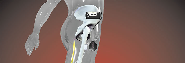
ActiGait
The system consists of a heel switch (3 in diagram above) which communicates wirelessly with a control unit (1), which is worn on a magnetic clip anywhere discreet as chosen by the patient. This unit allows the patient to adjust the intensity of the stimulation. An electromagnetic signal is painlessly sent through the skin at the upper thigh via a lightweight antenna (2) to the implant (4), which converts that signal into electric current for the 4 channel electrode cuff positioned around the Common Peroneal Nerve. The four channels can be programmed to allow for selective nerve bundle stimulation and balanced dorsiflexion / eversion.
Review ActiGait vs StimuStep
A clinical follow-up (a mixed population of 46 cases since 2006 of which 42 were reviewed for the study) of StimuStep was presented by Taylor20,21. The StimuStep users were selected from existing surface FES users. Reasons for selection of the implant were skin irritation, patients’ difficulties with electrode placement or anticipated long term use. Indications were stroke (18 cases), MS (17 cases, 1 bilateral), traumatic head injury (3), incomplete spinal cord injury (2), brain tumour (1), Parkinson’s (1), transverse myelitis (2) and cerebral palsy (1). 4 patients were not followed up due to : 2 non-functioning implants, 1 explantation because of infection and 1 for poor response because of abnormal nerve anatomy. The main benefits to patients reported were improvements in walking speed (18%) and a three-minute walking distance (23%).
Complications were reported as 6 electrode failures, 9 cases of nerve dysfunction (likely due to epineural electrode positioning and direct pressure on the receiver). Electrical sensation only improved 1 point out of 10 in comparison to surface stimulation with two cases even more uncomfortable level of sensation than surface stimulation. Five cases of skin reaction were reported. The patient still needs to wear a cuff directly on the skin which has a large contact area and some contact pressure. Despite implanted device patients still experienced issues with electrode and control box position. 11 of the first 16 cases also had reliability problems with the stimulation channel to the superficial branch of the nerve.
ActiGait was the subject of a safety and performance study conducted in three centres in Denmark1, which established safety using nerve conduction velocity and performance improvements in walking speed (20%) and distance walked in four minutes (14%). Long-term improvements were detected in walking speed and distance when stimulated, and the orthotic effect of stimulation showed statistically significant improvement. Furthermore, qualitative responses highlighted improvement in confidence with less fear of falling, promoting the long-term potential to provide a positive effect on personal well-being, safety and performance1,8. Similar patient benefits were reported in a more recent study22 showing a 24.5% increase in walking speed, and 17% increase in walking distance in the six-minute walking test. In addition to walking speed and endurance, the kinematic and biomechanical changes were investigated in five subjects by Ernst et al23. The study demonstrated a restored ankle joint movement towards a more physiological pattern as seen in normal gait.
The ActiGait implant complication rate was followed up by the manufacturer’s internal quality control24. Since the introduction of the newest revision of the device in February 2011, 115 implantations were reviewed (mixed population, indication stroke). All reported complications had been operator-caused (surgical procedure / general surgical risk), none have been caused by the implant. The complications reported were 4 cases of infection and 4 cases of temporary nerve damage (3 of which are functional with ActiGait in situ at the time of writing). Surgical reasons were established as causes for those nerve damages. Since the last revision of the surgical procedure 60 implantations have been carried out with no nerve damages, no implant failures nor incidents with external components. For licensing reasons the main indication for ActiGait is stroke. However, first implantations have been carried out for alternative indications in our practice, such as head injury and multiple scleroris with very good individual outcomes in the improvement of quality of life. Further work will be undertaken in implantations for other indications.
It appears that by direct comparison the benefits of both implants are compatible. However, comparing the reported complications, ActiGait has despite higher case numbers a lower complications rate, none of which are caused by the device itself.
References
1.Burridge JH, Haugland M, Larsen B, Pickering RM, Svaneborg N, Iversen HK, et al. Phase II trial to evaluate the ActiGait implanted drop-foot stimulator in established hemiplegia. Journal of Rehabilitation Medicine, 2007;39(3):212-8.
2.Burridge J, Taylor P, Hagan S, Wood D, Swain I. The effect of common peroneal nerve stimulation on quadriceps spasticity in hemiplegia. Physiotherapy, 1997;83(2):82-9.
3.National Institute for Health and Clinical Excellence. Interventional procedures overview 278: Functional electrical stimulation for drop foot of central neurological origin. 2009.
4.National Institute for Health and Clinical Excellence. Clinical guideline 8: Multiple Sclerosis; National clinical guideline for diagnosis and management in primary and secondary care. 2004.
5.Wade D. Measurement in Neurological Rehabilitation. Oxford, UK: Oxford University Press; 1992.
6.Kottink AI, IJzerman MJ, Groothuis-Oudshoorn CG, Hermens HJ. Measuring Quality of Life in Stroke Subjects Receiving an Implanted Neural Prosthesis for Drop Foot. Artificial Organs, 2010;34(5):366-76.
7.Barrett C, Taylor P. The Effects of the Odstock Drop Foot Stimulator on Perceived Quality of Life for People With Stroke and Multiple Sclerosis. Neuromodulation: Technology At The Neural Interface. 2010;13(1):58-64.
8.Burridge JH, Haugland M, Larsen B, Pickering RM, Svaneborg N, Iversen HK, et al. Patients’ perceptions of the benefits and problems of using the ActiGait implanted drop foot stimulator. Journal of Rehabilitation Medicine. 2008;40:873-5.
9.Department of Health. National Stroke Strategy. 2007.
10.Department of Health. National Service Framework for long term conditions. 2005.
11.CEP10012: Centre for evidence-based Purchasing. Economic report: functional electrical stimulation for drop foot of central neurological origin. NHS Purchasing and Supply Agency. 2010. London.
12.Intercollegiate Stroke Working Party. National Clinical Guideline for Stroke, 4th edition London: Royal College of Physicians 2012. ISBN 9781860164927
13.CEP10010: Centre for evidence-based Purchasing. Buyer’s guide: functional electrical stimulation for drop foot of central neurological origin. NHS Purchasing and Supply Agency. 2010. London.
14.Mann GE, Jolley CL, Taylor PN. An Investigation into the effect of functional electrical stimulation on mobility and quality of life in patients with multiple sclerosis. Proceedings of the 10th Annual Conference of the International FES Society. July 2005, Canada.
15.Daly J, Roenigk K, Holcomb J, Rogers JM, Butler K, Gansen J, McCabe J, Fredrickson E, Marsolais E, Ruff R. A randomized controlled trial of functional neuromuscular stimulation in chronic stroke subjects. Stroke, 2006, 37(1):172-8.
16.van Swigchem R, Weerdesteyn V, van Duijnhoven HJ, den Boer J, Beems T, Geurts AC. Near-Normal Gait Pattern With Peroneal Electrical Stimulation as a Neuroprosthesis in the Chronic Phase of Stroke: A Case Report. Arch Phys Med Rehabil, 2011, 92:320-4.
17.Waters RL, McNeal D, Perry J. Experimental correction of foot drop by electrical stimulation of the peroneal nerve. Journal of Bone Joint Surgery Am, 1975, 57:1047-1054.
18.Taylor PN, Burridge JH, Dunkerley AL, Lamb A, Wood DE, Norton JA, Swain ID. Patients’ perceptions of the Odstock Dropped Foot Stimulator (ODFS). Clinical Rehabilitation, 1999, 13:439-446.
19.Otto Bock Healthcare, Internal Report: MyGait Field Test Results, April 2013.
20. Taylor P, Wilkinson IH, Humphreys L, Kwan Y, Slade-Sharman D, Khan M, Hobby J. Clinical Experience of the STIMuSTEP Implated Dropped Foot Stimulator. International IFESS Conference 2012, Banff, Alberta, Canada.
21.Taylor P, Wilkinson I, Samuel V et al. A comparison of external and implanted EFS for correction of dropped foot. An audit of the STIMuSTEP service in Salisbury. 4th Annual UKRI IFESS Conference 2013.
22.Rohde V, Wachter D, Ernst J, Liebetanz D. Perneal stimulation for foot drop management after chronic stroke: Experience in 25 Patients, 63. Jahrestagung der Deutschen Gesellschaft fuer Neurochirurgie, 2012, Leipzig.
23.Ernst J, Grundeya J, Hewitta M, von Lewinskia F, Kaus J, Schmalz T, Rohde V, Liebetanz D. Towards physiological ankle movements with the ActiGait implantable drop foot stimulator in chronic stroke. Restorative Neurology and Neuroscience. In Press.
24.Statement about ActiGait Complication Rates by Dr. Andreas Hahn (Managing Director, nStim Services) 16 April 2013.
Sign up to receive our email newsletter with links to the latest content. ACNR is free, thanks to the support of advertisers. The editorial content is peer reviewed and remains completely independent unless clearly specified.